Dissertation on Nanoparticle Efficiency in Chemotherapy
Info: 7516 words (30 pages) Dissertation
Published: 17th Nov 2021
SYNTHESIS AND MANUFACTURING OF CONTROLLED AND TARGETED COAXIAL CYTOSTATIC POLYMER NANOPARTICLES THROUGH THE PROCESS OF ELECTROSPRAYING FOR DRUG DELIVERY SYSTEM
Abstract
In this study, the effects of changing the parameters for electrospraying process to efficiently fabricate nanoparticles with uniformity, spherical shape, and desired size of 20-50 nm range are elucidated. Probe sonication method with sonication time of 120 minutes was shown to be the preferred time to break down the molecules to achieve the nanoparticles with the lowest size. In all, it is shown that it is of utmost importance to carefully investigate how sonication, flow rate, tip to collector distance, amplitude relating to the intensity of the sonication, and applied voltage influences the sizes of the nanoparticles to further reduce the size of the particles to reach the desired size range.
INDEX WORDS: Electrospraying, Nanoparticles, Sonication.
Click to expand Table of Contents
Table of Contents
LIST OF SYMBOLS…………………………………………………………………7
LIST OF TABLES……………………………………………………………………8
LIST OF FIGURES………………………………………………………………..9
1 INTRODUCTION………………………………………………………….……..10
- Introduction to Malignancy……………………………………………10
- Types of Chemotherapy Treatments……………………………….11
- Cancer Statistics………………………………………………………12
- Chemotherapy Procedure……………………….……………………12
2 LITERATURE REVIEW…………………………………….………………14
2.1 Introduction to Polymers………………………………………………..14
2.2 Electrohydrodynamic Atomization……………………………..………15
2.3 Electrospinning of Nanofibers………………………………….….……15
2.4 Electrospraying of Nanoparticles……………………………………….17
2.5 Coaxial Process for Electrospraying……………………………………17
2.6 Introduction to BEHLKE Switch…………………………………….…19
2.7 Similar Techniques……………………………………………………..21
3 METHODOLOGY……………………………….………………….………23
3.1 Introduction………………………………………………………………..23
3.2 Model Design and Fabrication…………………………………………..23
3.3 Equipment for Preparation of the Solution………………………………23
3.4 Experimental Method for Preparing the Solution……………………….24
3.5 Sonication Process……………………………………………………….24
3.6 Experimental Method and Equipment for Electrospraying……………..25
3.7 Testing of BEHLKE Switch……………………………………………….27
3.8 Summary of Analysis………………………………………………………27
3.9 Criteria for Success………………………………………………………28
4 FINDINGS OF THE STUDY……………………………………………….29
4.1 Introduction………………..………………………………………………29
4.2 Results for Trials with Sonication Time as Varying Parameter………29
4.3 Results for Trials with Voltage, Sonication Time, Flow Rate, Tip to
Collector Distance as Varying Parameter……31
4.4 Results for BEHLKE Switch………………………………………………32
5 CONCLUSIONS AND RECOMMENDATIONS…………………………..33
5.1 Summary of Work …………………………………………………………33
5.2 Conclusion………………………………………………………………….33
5.3 Suggestions for future work………………………………………………34
REFERENCES…………………………………………………………………35
List of Symbols
| ρ | Density (kg/m3) |
| l, d | Distance (mm) |
| g | Gravity (m/s2) |
| m | Mass (g) |
| n | Mole (mol) |
| ∈ | Permeability Factor (m2) |
| R, r | Radius (mm) |
| ѵ | Speed (mm/s) |
| γ | Surface Tension (N/m) |
| t | Time (s) |
| V | Voltage (kV) |
| v | Volume (ml) |
List of Tables
Table 3.1. Experimental Trials with Sonication as Varying Parameter
Table 3.2. Experimental Trials with Sonication, Voltage, Distance, and flow rate and Varying Parameters
Table 3.3 Electrospinning Machine Parameters
List of Figures
Figure 2.1. Basic Schematic of Electrospinning (Wu, Yiquan, and Robert Clark, 2008)
Figure 2.2. Effects of Process Parameters on Nanofibers and Nanoparticles (Zamani, Maedeh , Prabhakaran, and Ramakrishna, 2013)
Figure 2.3. Basic Schematic of Electrospraying (Wu, Yiquan, and Robert Clark, 2008)
Figure 2.4. Schematic of Electrohydrodynamic Atomization Process (Marjan Enayati, Zeeshan Ahmad, Eleanor Stride, and Mohan Edirisinghe, 2009)
Figure 2.5. Illustration of Printed Wiring Information for BEHLKE Switch (BEHLKE Catalog C8, 2016)
Figure 2.6. Illustration of Printed Wiring Information for BEHLKE Pump (BEHLKE Catalog C8, 2016)
Figure 2.7. Illustration of Printed Wiring Information for BEHLKE Fan (BEHLKE Catalog C8, 2016)
Figure 4.1. Results for Trials with sonication time as varying parameter
Figure 4.2 Results for Trials with Voltage, Sonication Time, Flow Rate, and Tip to Collector Distance as Varying Parameters for Trial 1 from Table 3.1
Figure 4.3 Results for Trials with Voltage, Sonication Time, Flow Rate, and Tip to Collector Distance as Varying Parameters for Trial 2 from Table 3.2
Chapter 1. Introduction
1.1 Introduction to Malignancy
Abnormal growth of cells that invade normal tissue can be either in benign or malignant forms. Malignancy is cancerous cells that can spread, invade, and destroy normal tissues. The growth and formation of malignant cells starts at molecular level and it is unpredictable due to the swift rate of expansion and uncontrollability of cells. The interaction between person’s genetic factors and external agents such as physical carcinogens, chemical carcinogens, and biological carcinogens are the factors that transform normal cells to benign tumors then ultimately to malignant cells (WHO, 2017).
Malignant cells do not die on their own, therefore, require treatments such as radiotherapy, surgery, hormonal therapy, biological therapy, or chemotherapy (Cancer Research UK, 2015). While cancer or tumors can be surgically removed for solid tumors, due to the abnormality in shapes and structures of the cells in most cases of cancer, it is not possible to entirely remove the tumors or cancer with surgery alone. The most common type of treatment removal of cancer is chemotherapy. Chemotherapy is a treatment method that is used to treat cancer using the drugs called cytostatics. There are four types of chemotherapy treatments that are utilized based on the final aim in removal of malignant cells: curative, adjuvant, neoadjuvant, and palliative chemotherapy (PubMed Health, 2016). The treatment options are dependent upon the condition of the patient. The conditions include, the type of cancer, the structure of cancer cells, whether if cancer has distributed to other tissues, and the health of the patient (Cancer Research UK, 2015).
1.2 Types of Chemotherapy treatments
The goal of curative chemotherapy is to completely remission all cancerous cells from the body for permanent cure. As mentioned above, surgery alone cannot remove cancer, when this is the case, adjuvant chemotherapy is the option. Adjuvant chemotherapy removes the cells that are left in the body after the removal of tumor or malignant cells through surgery. The goal of adjuvant chemotherapy is to prevent malignant cells from growing back. Next, for cases of tumors with large mass, neoadjuvant chemotherapy is the option. When it is not possible to remove large tumors with surgery, neoadjuvant chemotherapy is administered to shrink the size of the tumor so that surgery can be an option to remove the tumor. This process not only makes it possible to operate on a tumor but also makes the surgery less invasive. The last type of chemotherapy treatment is palliative. Palliative chemotherapy is useful when removal of tumor is no longer an option or impossible. Palliative chemotherapy temporarily slows down the disease and relieves the symptoms and improves the patient’s quality of life, however, it does not provide a permanent solution for the removal of malignant cells. (Cancer Research UK, 2015)
1.3 Cancer Statistics
According to the World Health Organization, one of the primary causes of death around the world is cancer with approximately 14 million cases as of 2012 (WHO, 2017). National Cancer Institute states that as of 2016 estimated 1,685,210 new cases of cancer are diagnosed in United States alone with a mortality number of 595,690 (National Cancer Institute, 2017). According to American Cancer Society, next to skin cancer, breast cancer is the most common type of cancer in the United States for the women with a chance of 1 in 8 women will develop breast cancer and an estimated number of 40,610 women will die of breast cancer in 2017 (The American Cancer Society, 2017). It is the third highest cancer related death after lung and pancreatic cancers. Lung and bronchus cancer, prostate cancer, colon and rectum cancer, bladder cancer, melanoma of the skin, non-Hodgkin lymphoma, thyroid cancer, kidney and renal pelvis cancer, leukemia, endometrial cancer, and pancreatic cancer are the most frequently diagnosed cancers in the United States with an estimated mortality of 155,870 and 43,090 for lung and pancreatic cancers, respectively (National Cancer Institute, 2017). The staggering cancer deaths also include deaths that are occurred due to treatment such as chemotherapy.
1.4 Chemotherapy Procedure
Chemotherapy can be administered to body by either infusing cytostatic drugs into the veins through needles or through tablets. Cytostatic drugs inhibit the cells from growing and dividing, therefore, stopping the cells from multiplying and spreading through the body. When cytostatic drugs are administered through infusion into veins, the drug travels through entire body if local chemotherapy treatment is not used, this process is vital to kill any cancerous cells that are not discovered during examination or that cannot be removed through either radiation or surgery.
As crucial as the infusion technique is, the negative side effect of cytostatic drug is that it destroys both good and bad cells when it travels throughout the body since the drug cannot differentiate between malignant, benign, or normal cells. Unfortunately, the normal cells that can be destroyed through cytostatic drugs include, “blood producing cells, hair cells, cells of mucous membranes of mouth and throat area and of the digestive system” (PubMed Health, 2016). And other adverse effects include, “hair loss, anemia, nausea, vomiting, diarrhea, and infections in mouth” (PubMed Health, 2016).
The dosage and type of drugs used to administer chemotherapy play a vital role in side effects experienced by patients. As non-localized chemotherapy creates more and more undesirable side effects for the patient’s body and localized chemotherapy is not ideal for all cancer types to inhibit malignant cells, nanoparticles come to the forefront as a potential source of controlled and targeted cytostatic polymer nanoparticle drug delivery system. If the treatment by controlled and targeted nano-encapsulated cytostatic polymer drug delivery system is used to administer in place of standard chemotherapy treatment procedure, then the number of vital cells destroyed during chemotherapy process will decline.
In order to achieve this, nanoparticles with the diameter range of less than 20-50 nanometers should be manufactured with the encapsulated drug. However, compared to the production of fibers, nanoparticles are more difficult produce in the desired scale range since the process is very unpredictable and achieving uniform size particles is a difficult process.
In order to achieve particles with desired size and uniformity, the parameters for electrospinning process should be studied to understand how the changes will affect the size, shape, uniformity of the particles. Therefore, if the parameters used for electrospinning are altered, then the particles in desired scale range can be achieved to deliver the cytostatic drug in molecular level.
Chapter 2. Literature Research
2.1 Introduction to polymers
A polymer is a specific kind of matter with uniform properties which contain sizable number of structural units joined by the same type of linkage, which often forms into chain like structures (Yu, 2011.) Compared to any other material, when polymers are broken into nanoscale, they have a dramatic increase in surface to volume ratio, flexibility in surface functionalities, and superior mechanical performance (Zamani, 2013).
Since the surface to volume ratio dramatically increases for polymers as the size approaches nanoscale, they are considered to be surface dominated materials, which rely on melting point, adhesion, and rate of reaction. The ratio of surface to volume increases the dipole interactions of the atoms changing the adhesion properties. Therefore, as the polymers size decreases, the adhesion properties increase making the material super absorbent, making it a good product for encapsulating cytostatic drugs.
To produce continuous nanofibers or nanoparticles, electrospinning and electrospraying process is used for production of large quantities of fibers or particles. Polymers can be used as biodegradable materials to encapsulate cytostatic drugs to gradually release through diffusion and degradation.
2.2 Electrohydrodynamic Atomization
Electrohydrodynamic atomization, a process by which electrostatic forces are used to produce fibers or particles of various sizes through fluid jet, can manufacture polymer nanofibers and nanoparticles by the two modes of electrohydrodynamic process known as electrospinning and electrospraying. These processes are capable of producing fibers and particles that can help in the process of controlled drug delivery encapsulation. Electrospraying and electrospinning are top down fabrication approaches with the classification of characteristic dimension of one dimensional for nanofibers and zero dimensional for nanoparticles.
2.3 Electrospinning of nanofibers
The popular method to generate nanofibers is by Electrospinning. During electrospinning, a strong electrical potential is applied to the polymer liquid, resulting in the accumulation of electrical charges on the surface of the liquid droplet on the tip of capillary. The Coulombic repulsion of the charges overcomes the surface tension of the polymer droplet at a critical voltage resulting in ejection of charged jet in conical shape from the tip of the droplet. The charged jet then accumulates the fibers as in Figure 2.1, onto the grounded electrode while evaporating the solvent. Electrospinning directly encapsulates the drugs into nanofibers. Due to the high surface tension and also the porosity of the fibers in three-dimensional scale, the rate at which drug diffuses is reduced, leading to an efficient drug release method (Zamani, 2013)
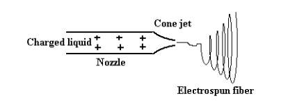
Figure 2.1. Basic Schematic of Electrospinning.
2.4 Electrospraying of nanoparticles
Another promising drug delivery method is the Electrospraying as shown in Figure 4, produces nanoparticles. The process of electrospraying is similar to that of electrospinning. The advantage of electrospraying particles in comparison to electrospinning of fibers for the administration of cytostatic drug is that, the fibers agglomerate with each other and intertwine with each other making the releasing of encapsulated drug at an uncontrolled rate.
While electrospraying is a method to manufacture nanoparticles, the parameters that transition the manufacturing process from electrospinning to electrospraying include the distance between the collector plate to the spinneret, density of the solution, surface tension of the polymer solution, gravitational force, the forces acting on the meniscus of the droplet in the capillary, the flow rate of the polymer solution, the applied voltage, and the permeability. When the properties of solution are altered and the parameters of the process are changed, the continuous and charged jet can be broken down into particles, resulting in particles of different size and shape as shown Figure 2.2 (Zamani, 2008).
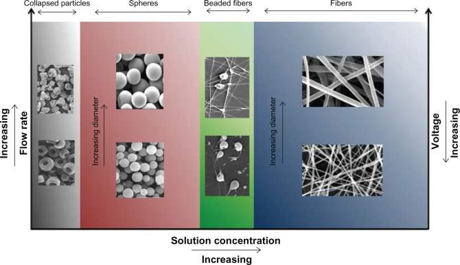
Figure 2.2. Effects of Process parameters on fibers and particles.
The main components of electrospraying are: high-voltage power supply, spinneret, and conductive plate as collector. The basic electrospraying process as shown in Figure 2.3, follows the key stages, starting with the generation of a droplet when high voltage power source is applied to the nozzle, followed by the formation of Taylor cone, and then of the launching of the jet, the jet is then elongated into a straight segment resulting into development of whipping instability and ultimately resulting in the solidification of the particles which is collected on the collector plate.
In more depth, monodisperse particles with uniform particle size can be achieved through electrospraying by placing a ring connected to the intermediate voltage below the spinneret, which stabilizes the electrospraying jet, as highly charged particles are strongly attracted by grounded or oppositely charged objects, uniform collection of particles can be achieved. Another possibility is by separating the particles according to their mass/surface-charge density.
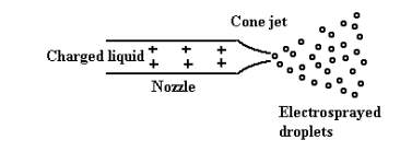
Figure 2.3.Basic Schematic of Electrospraying.
2.5 Coaxial Process for Electrospraying
To encapsulate the drugs, coaxial process with two solutions flowing through a coaxial capillary is used to produce coaxial structures (Wu,Clark, 2008). The basic schematic of electrohydrodynamic atomization process of electrospraying of coaxial polymer and drug solution is shown in Figure 2.4.
Figure 2.4 Schematic of Electrohydrodynamic Atomization process
For coaxial electrospinning process, a smaller diameter capillary tube is concentrically connected into a larger diameter capillary tube for coaxial configuration. Then, the connected capillaries are attached to an independent reservoir that consists of syringe pump, which aids in releasing the solution at a specified flow rate through the coaxial cone to be collected onto the grounded electrode as shown in Figure 2.4. The purpose of the controlled drug delivery is to deliver and retain sufficient amount of drug for an adequate period of time through controlling the rate, time, and location of drug release (Zamani, 2008).
2.6 Introduction to BEHLKE switch
During the electrospraying process, it is important to achieve a Taylor cone. A Taylor cone can form when a fluid with electric conductivity is exposed to an electric field. The threshold voltage in this electric field allows the liquid to break away from the normal surface tension, creating the cone structure with a round tip. A nanoparticle encapsulating the cancer drug is then coaxially spun onto the grounded collector continuously to form layers of drug encapsulated particles. However, the next step in achieving smaller and more consistent nanoparticles is to electrically pulsate the voltage in the testing environment.
A BEHLKE switch as shown in Figure 2.5, is a fast-high voltage, variable on time, push pull switch will be incorporated into both electrospinning and electrospraying processes. Push pull switches have high rise and fall times. Rise time, is the time required for a pulse to rise from 10 percent to 90 percent of its steady value. Rise time can easily be measured on an oscilloscope and can be applied to any linear network (Rise Time, 2005). Rise time is taken by signal to change from a specified low value to a specified high value when describing either voltage and current step function. Inversely, fall time or pulse decay time, is time required for the amplitude of pulse to decrease from a specified high value which is 90 percent of the peak value to another specified low value which is 10 percent of the peak value (Fall Time, 1996). Since, it is a high voltage push pull switch, it has high frequency (Felger, 2017).
Figure 2.5. Illustration of Printed Wiring Information.
According to BEHLKE’s website, fast push pull switches are sensitive for reverse currents from inductive load or form increased wiring inductance. “Reverse currents may turn on the parasitic MOSFET diodes in an undefined way, which leads to short circuit within the switching paths, ultimately resulting in failure. Therefore, it is imperative to make sure that no current swings back into the output of the switch. To make sure this problem does not occur, an oscilloscope can be used to measure the maximum 10 percent of the actual operating voltage” (BEHLKE, 2016).
This high frequency push pull switch will pulse on and off nearly every 100 nanoseconds. The pulsing at this speed should produce more uniform nanofibers and nanoparticles that fall within the tolerances needed for the desired application. The high frequency push pull switch will ideally cut the nanofibers and nanoparticles every time the switch is off. To electrospin the nanofibers and electrospray the nanoparticles, a DC voltage source must be used. For the testing equipment used, this voltage needs to be between 20-30kV. The frequency that the BEHLKE switch emits may have an impact on the mass flow rates of the two solutions, thus potentially the need to readjust the two flow rates. In order to incorporate the switch into the electrospinning machine, a pump as shown in Figure 2.6, a fan as shown in Figure 2.7, required resistors , capacitors, and two controllers to control the frequency of the switch and pump are to be wired together (Felger, 2017).
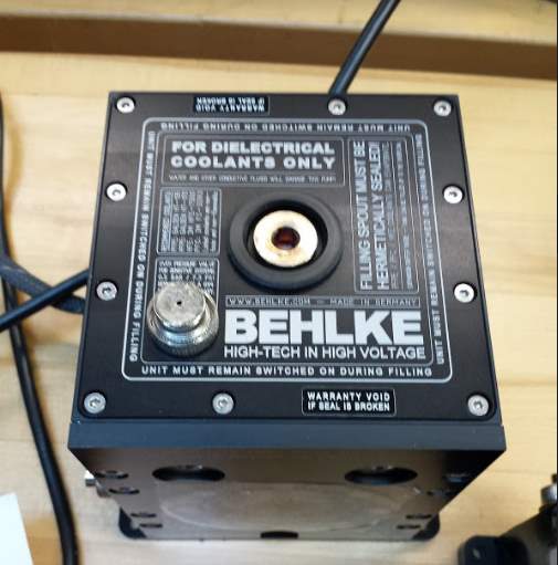
Figure 2.6 Illustration of Printed wiring information for pump
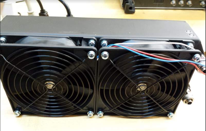
Figure 2.7 The fan that is to incorporated into the electrospinning machine
Prior to placing the mentioned devices into the electrospinning machine, it is vital to make sure the switch is functioning so that the switch would not damage the electrospinning machine.
2.7 Similar Research/Techniques
Another technique that is used to fabricate to nanoparticles is the process of emulsion. For the emulsion process, Molecules are dispersed or dissolved into a polymer solution and emulsified to form micro-droplets that are further dried after solvent removal (Bock, 2011) The disadvantage of emulsion technique is that since organic solvents are used, if not carefully controlled, the protein based drugs can be denaturized resulting in inconsistent encapsulation of the drug. In addition, the size of the particles produced through this process inhomogeneous and broad, contributing to their lack of reproducibility (Bock, 2011) Research conducted by Bock, tested the size of nanoparticles with various set of parameters for each condition. The generated particles were in 10-17 micro-range.
CHAPTER 3. METHODOLOGY
3.1 Introduction
This section of the thesis covers procedures for the experimental studies. An electrospinning machine is used for electrospraying for the experimental portion of the study. At various parameters tested, the sizes of the particles are examined under the scanning electron microscope to determine how the parameters affect the size, shape, and the uniformity of the particles.
3.2 Model design and fabrication
Based on Saqib Iqbal’s research, “Encapsulation of Anticancer drugs (5-Fluorouracil and Paclitaxel) into Polycaprolactone (PCL) Nanofibers and in vitro testing for sustained and targeted therapy” the polymer solvent made of formic acid, acetic acid, trifluoroethanol with the solute of polycaprolactone resulted in the lowest diameter for the nanofibers, therefore, the same solution mixture with the same compositions for each material was tested in the study for producing nanoparticles (Iqbal, 2016). For each trial, the solution was made using the same concentration of material but the particles were examined after changing the parameters.
3.3 Equipment for the preparation of the solution
The Georgia Southern’s nanomaterial laboratory is equipped electrospinning machine for the fabrication of nanoparticles. However, prior to fabrication, the solution has to be prepared and then sonicated. The materials required for the preparation of the solution are three pipets, magnetic hot plate and stirring machine, weight machine, beakers, sonication machine, weigh boat, and parafilm tape.
3.4 Experimental Method for preparing of the solution
It is imperative to follow the rules and guidelines of the laboratory. Therefore, prior to making the solution, it is crucial to wear the lab coat, goggles, and latex gloves to handle hazardous materials.
First, obtain three, 10 ml pipette to obtain acetic acid, formic acid, and trifluoroethanol (TFE). The concentration of the acids to be used for each trial are 47.5% acetic acid, 47.5% formic acid, and 5% TFE. A solvent of 20 ml is to be made. To make a solvent of 20ml, 9.5ml of formic acid, 9.5 ml of acetic acid, and 1 ml TFE is required. It is crucial to use three different pipettes to obtain the acids. When desired amount of each acid is obtained, the acids can be placed in a single 50ml beaker.
The next step is to obtain 140 mg/ml of polycaprolactone (PCL) which is 2.8g of PCL for 20 ml of solvent. Place a weigh boat on the weight machine and zero the machine. Once the machine is zeroed, measure PCL of 2.8g. Place the beaker containing the solvent on the magnetic hot plate and stirring machine with magnetic stirring bar placed inside of the beaker. Cover the beaker with parafilm tape and slowly increase the speed of the machine until optimum speed is reached. This is done to make sure all the acids are mixed properly before adding PCL.
After the solvent is stirred for 5 minutes, add PCL in small quantities to the solvent, place the parafilm foil, turn on the stirring machine. PCL dissolves very slowly at room temperature, therefore, it was mixed on hot plate.
Follow this process to dissolve the entire solute in the solvent. The mixed solution is now ready for sonication.
3.5 Sonication Process
Sonication is a method that is used to break down molecules in smaller size by applying sound energy to vibrate the particles in a sample through a probe. For the first set of experiment, set the amplitude of the probe to 20. Prior sonicating the solution, cover the beaker with parafilm foil to protect the solution from evaporating and also place the machine under fume hood to limit exposure to hazardous fumes.
As the amplitude of the sonicator is increased, the volatility of the solution increases, so it is crucial to cover the solution with parafilm tape. The bubbles that are seen in the solution while it is being sonicated, are due to cavitation that results in tiny shockwaves that break the cells apart. For the first trial, the solution is sonicated for a total time of fifteen minutes. Tables 3.1 and 3.2 show the sonication time for next set of trials. The electrospinning machine parameters as shown in Table 3.3 are set at same values for all the trials.
The solution is now ready for electrospraying. It is vital to understand how changing the parameters for electrospraying affect the particles, therefore, it is important to first fabricate nanoparticles with just the polymer solution. Once, the desired size, shape of the particles are achieved through a specific change in parameter, use that parameter as a base to fabricate coaxially encapsulated nanoparticles.
3.6 Experimental Method and Equipment for Electrospraying
Electrospinning machine is a high voltage machine; therefore, it should be used cautiously by following all procedures. Start the machine by turning on the power source, make sure a dehumidifier is attached to the machine in order to reduce the level of humidity in the testing environment. The polymer solution needs to be placed in a 6-ml syringe, and then should be secured to feed pump 1. Since only polymer solution is being electrosprayed, pump 2 will not be utilized for this experiment. Place an aluminum foil covering the surface of the collector plate to collect the sample electrosprayed particles. Secure the testing environment by closing and locking the equipment door.
Once the fabrication process is in progress, the door cannot be opened due to the high voltage in the chamber. In order to open the door, the experiment has to be stopped or paused. Once the door is secured, change the parameters for tip to collector distance, flow rate, and applied voltage on the touch pad located on the equipment to the values shown in Tables 3.1, 3.2, and 3.3. Once all the parameters are set, first select preset and then press start to start the fabrication process.
Table 3.1. Experimental Trials with sonication as varying parameter.
| Solvent | Trial | Solvent (ml) | Solute (g) | Sonication time (min) | Amplitude (%) | Flow rate (m//hr) | Applied Voltage (kV) | Tip to Collector distance (mm) |
| 47.5% FA, 47.5% AA, 5% TFE# | 1 | 20 | 2.801 | 60 | 20 | 0.7 | 28 | 145 |
| 2 | 20 | 2.801 | 90 | 20 | 0.7 | 28 | 145 | |
| 3 | 20 | 2.803 | 105 | 20 | 0.7 | 28 | 145 | |
| 4 | 20 | 2.803 | 120 | 20 | 0.7 | 28 | 145 |
Table 3.2. Experimental Trials with varying parameters
| Solvent | Trial | Solvent (ml) | Solute (g) | Sonication time (min) | Amplitude (%) | Flow rate (m//hr) | Applied Voltage (kV) | Tip to Collector distance (mm) |
| 47.5% FA, 47.5% AA, 5% TFE# | 1 | 20 | 2.798 | 100* | 30 | 0.6 | 30 | 135 |
| 2 | 20 | 2.8 | 120 | 30 | 0.7** | 30 | 135 | |
| 3 | 20 | 2.8 | 150 | 30 | 1 | 34 | 120 | |
| 4 | 20 | 2.8 | 150 | 30 | 1 | 36 | 120 | |
| 5 | 20 | 2.8 | 150 | 30 | 1 | 36 | 110 | |
| 6 | 20 | 2.8 | 150 | 30 | 1.5 | 36 | 100 |
Table 3.3. Electrospinning Machine Parameters
| Transverse speed (mm/s) | Transverse length (mm) | Rolling speed
(mm/s) |
Rotation Speed (mm/s) |
| 10 | 100 | 0.33 | 0 |
3.7 Testing of BEHLKE Switch
In order to incorporate the BEKLKE switch into the electrospinning machine, it is first tested. A testing circuit consisting of the switch, low voltage source, function generator, oscilloscope, and function generator were used. Function generator controlled the frequency, ultimately controlling the on and off times for the switch, oscilloscope was used to observe the time interval and voltage in waveform with constant voltage of 15V, the frequency on the function generator was set to 2kHz with an amplitude of 5V. The BEHLKE Switch was tested by incorporating the switch with a pump- fan cooling system.
3.8 Summary of Analysis
Once the samples are collected, analysis must be done to compare the sizes and structures of nanoparticles to see which change in parameter gives the best results for the size, shape, and uniformity of the particles. In order to analyze the sample, it must be examined under scanning electron microscope. By analyzing the samples, they can be compared to other similar research and to Figure 2.3 for the validation of the experiment. Also, it has to be noted that the values in Table two marked with * and ** indicate that most of the solution was evaporated during the sonication time of 100 minutes, leaving the solution with 2 ml at the end of 100 minutes and the flow rate was changed from 0.6 ml/hr. to 0.7 ml/hr. because the machine was not electrospraying but was electrospinning at 0.6 ml/hr.
3.9 Criteria for Success
When planning on electrospraying a solution, it is important to use the solution while it is newly made. Even though the solution can be placed in the refrigerator and be used later, the best results are shown when the solution was used when right after it was made. If the solution is refrigerated, the solution becomes condensed and if was already sonicated then the effects of sonication process will wear out so, it would again have to be heated, stirred, and sonicated. Doing this will does not give a true value for the change in parameters.
CHAPTER 4. FINDINGS OF THE STUDY
4.1 Introduction
This chapter covers the results and discussion of the experimental analysis carried out in the research. The information is organized as follows:
- First, fabrication process was carried out with only sonication as the varying parameter to verify if increasing sonication time decreases the size of the particles produced.
- After the nanoparticles are observed and analyzed under scanning electron microscope for varying sonication time, the parameters for applied voltage, flow rate, tip to collector distance, and again sonication time were changed to see how the nanoparticles are affected.
4.2 Results for Trials with sonication time as varying parameter
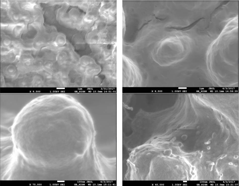
Figure 4.1. Top left is trial 1, top right is trial 2, bottom left is trial 3, and bottom right is trial 4.
Based on the results from SEM, it is identified that increasing sonication time resulted in the bonds to be broken down, therefore, resulting in in particles with smallest size as in trial 4 as shown in Figure 4.1.
In trial one, light interference from the scanning electron microscope blurs the picture but it can be seen that the bonds are not broken down, there is still sign of agglomeration, with particles in micrometer scale.
When the sonication time was increased to 90 minutes for trial 2, individual particles started to form, the particles even though are still at micrometer range, they are less agglomerated.
The sonication time was further increased to 105 minutes and then finally to 120 minutes for trials 3 and 4, respectively.
As seen in Figure 4.1, the trial with 120 minutes of sonication produced particles that are in nanometer range and those particles were smaller in size compared to trial 3. For 120 minutes of sonication resulted in particles in nanometer range, 120 minutes was used as the base time for the amount of sonication as shown in Table 2.1.
4.3 Results for Trials with voltage, sonication time, flow rate, and tip to collector distance as varying parameters
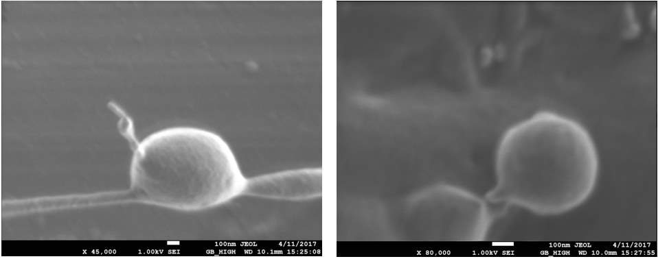
Figure 4.2 shows the results for trial 1 from Table 2.2.
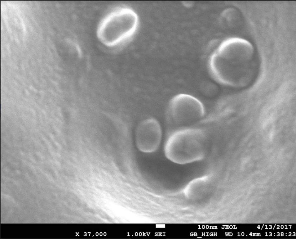
Figure 4.3 Trial 2 from Table 2.3
Figures 4.2 and 4.3 shows several particles in nanometer range and the size is decreasing with increasing of sonication and by increase in voltage applied. The tip to collector distance was also decreased in these trials, however, it is not how the change distance might have affected the size, therefore, the next set of trials will be performed with just changing the distance between tip to collector plate. The next set of trials with be conducted to examine how changing only one parameter at a time affects the size of the particles to further examine how changing all properties based on the best results from each varying parameter affects the size.
4.4 Results for BEHLKE Switch
The initial experiment of the switch with function generator, oscilloscope, and a constant voltage source of 15V, resulted in a graph of instantaneous voltage signal on the oscilloscope as a function of time. The wave signal was a square wave with amplitude of 5V with time span of 50ms on and 50ms off. This experiment confirmed that the switch was operating.
Chapter 5. Conclusions and Recommendations
5.1 Summary of work
This research investigates how change in sonication times, applied voltage, tip to collector distance, flow rate of the solution can affect fabrication of nanoparticles produced by the method electrospinning. The solution mixture with 47.5% acetic acid, 47.5% formic acid, 5% TFE, and 140 g/ml PCL solute was analyzed under scanning electron microscope to see how the changes in parameters changed the size, shape, and uniformity of the particles.
5.2 Conclusions
The following conclusions are drawn from the study:
- Increasing the sonication time breaks the bonds and molecules resulting in less agglomeration of the particles and making the particles more uniform and spherical.
- Increasing the sonication time resulted in nanoparticles with decreasing size.
- As applied voltage, tip to collector distance, flow rate, and sonication time are all changed, the particles also decreased in size, however, further test should be conducted to see how each parameter affects the size.
- The BEHLKE switch when tested with a circuit consisting of oscilloscope, a constant 15V power source, and a function generator produced a wave signal of 5V, showing that the switch is operating.
5.3 Suggestions for future work
Based on the results of this thesis, it is recommended that each parameter should be tested individually to see the effects on the particles and then the best results from those trials should be combined to see the effects on the particles. Also, based on the functionality of the switch after being tested, it can be tested using high voltage source along with a cooling system to keep the components inside the switch from burning. If the switch works using the high voltage source, it can therefore be incorporated into the electrospinning machine.
REFERENCES
“BEHLKE Catalog C8.” BEHLKE Catalog C8. N.p., 05 Aug. 2016. Accessed November 5, 2017. http://www.behlke.com/separations/separation_c8.htm>.
Bock Nathalie, Woodruff Maira, Hutmacher Dietmar, Dargaville Tim. “Electrospraying, a Reproducible Method for Production of Polymeric Microspheres for Biomedical Applications.” Polymer, no. 3 (2011): 131-149. Accessed November 5, 2017.
Cancer Research UK. “General Cancer Information.” January 05, 2015. Accessed November 1, 2017. http://www.cancerresearchuk.org/about-cancer/cancer-in-general/treatment/chemotherapy/what-chemotherapy-is
“Fall Time.” Definition: Fall Time. N.p., 23 Oct. 1996. Accessed November 5, 2017.
Felger, Bethapudi. “Synthesis and Manufacturing of coaxial drug loaded nanostructure.” Nanomaterials Laboratory, Georgia Southern Univeristy. Accessed Novemeber 5, 2017.
Iqbal, Sakib, Rashid Mohammad, Arbab Ali. “Encapsulation of Anticancer drugs (5-Fluorouracil and Paclitaxel) into Polycaprolactone(PCL) Nanofibers and in vitro testing for sustained and targeted therapy.” (Dec 19,2016). Accessed November 5, 2017. Keywords: Nanofibers, Paclitaxel, 5 Fluorouracil, Encapsulation, Electrospinning, Cytotoxicity.
Marjan Enayati, Zeeshan Ahmad, Eleanor Stride, and Mohan Edirisinghe. “One-step Electrohydrodynamic Production of Drug-loaded Micro- and Nanoparticles.” Journal of The Royal Society Interface. The Royal Society Publishing, 14 Oct. 2009. Accessed November 5,2017.
National Cancer Institute. “Cancer Statistics.” March 22, 2017. Accessed November 2, 2017. https://www.cancer.gov/about-cancer/understanding/statistics
National Cancer Institute. “Cancer Types.” February 13,2017. Accessed October 31, 2017. https://www.cancer.gov/types/common-cancers
PubMed Health. “How does chemotherapy work?” January 14, 2016. Accessed November 1, 2017. https://www.ncbi.nlm.nih.gov/pubmedhealth/PMH0072611/.
“Rise Time.” Van Nostrand’s Scientific Encyclopedia (2005): n. pag. Web. Accessed November 5, 2017. http://eent3.lsbu.ac.uk/staff/klimop/RF/Rise%20Time.pdf>.
The American Cancer Society. “About Breast Cancer.” September 21, 2017. Accessed October 31, 2017. https://www.cancer.org/cancer/breast-cancer/about/what-is-breast-cancer.html.
World Health Organization (WHO). “Cancer.” February 2017. Accessed November 5, 2017. http://www.who.int/mediacentre/factsheets/fs297/en/.
Wu, Yiquan, and Robert Clark. “Electrohydrodynamic Atomization: A versatile proces for preparing materials for biomedical applications.” J. Biomater. Sci. Polymer 19, no. 5 (2008): 573-601. Accessed November 5, 2017. Keyword: Electrospraying; electrospinning; biomaterials; biomedical application; tissue engineering; drug delivery.
“Yflow® Coaxial Electrospinning & Electrospray Tech.” Yflow S.D. N.p., 20 Feb. 2015. Web. Accessed November 5, 2017. http://www.yflow.com/coaxial-electrospinning-electrospray-microencapsulation/>
Yu, G., Branford White, N.P. Chatterton, L.M. Zhu, L.Y. Huang, and B. Wang. “A Modified Coaxial Electrospinning for Prreparing Fibers from a High Concentration Polymer Solution.” EExpress Polymer Letters 5, no. 8 (2011): 732-41. Accessed November 5, 2017. Keyword: processing technologies, coaxial electrospinning, polymer fibers, productivity.
Zamani, Maedeh , Molamma Prabhakaran, and Seeram Ramakrishna. “Advances in Drug Delivery via Electrospun and Electrosprayed nanomaterials.” International Journal of Nanomedicine 8 (August 9, 2013): 2997-3017. Accessed November 5, 2017. Keyword: electrospinning, electrospraying, gene delivery, growth-factor delivery, cancer therapy, wound dressing.
Zheng-Ming Huang, Y. Zhang , M. Kotaki and S. Ramakrishna. “A review on polymer nanofiber by electorspinning and their applications in nanocomposites.” Elsevier Journal (April 8, 2003). Accessed November 5, 2017. Keyword: electrospinning.
Cite This Work
To export a reference to this article please select a referencing stye below:
Related Services
View all


Related Content
All TagsContent relating to: "Cancer"
Cancer is a disease in which cells grow or reproduce abnormally or uncontrollably. Cancerous cells have the potential to spread to other areas of the body in a process called metastasis.
Related Articles

DMCA / Removal Request
If you are the original writer of this dissertation and no longer wish to have your work published on the UKDiss.com website then please: