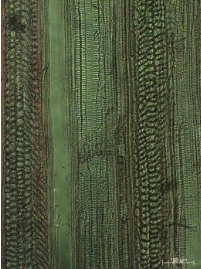
Anatomical Properties of Nine Indigenous Rattan Species of Jambi, Indonesia
Info: 6293 words (25 pages) Dissertation
Published: 9th Dec 2019
ANATOMICAL PROPERTIES OF NINE INDIGENOUS RATTAN SPECIES OF JAMBI, INDONESIA
Various rattan species grows naturally in Jambi, Indonesia, i.e. opon (Plectocomiopsis geminiflora (Griff.) Beccari), udang (Korthalsia flagelaris Miquel), getah (Daemonorops micracantha (Griff.) Beccari), duduk (D. didymophylla Beccari), tunggal (Calamus laevigatus Martius), sijau (C. tumidus Furtado), buruk ati (C. insignis Griff. var. longispinosus Dransfield), batu (C. zonatus Beccari), and paku (C. exillis Griff.). The rattan species are classified as lesser known species, which its properties are largely unknown to rattan supplier and consumers. This paper observes the anatomical properties of nine indigeneous rattan species of Jambi. Anatomical observations were conducted from solid, sectioned and macerated samples. Results show that anatomical properties become a diagnostic characteristic for rattan species identification and specific characteristic has been developed for key species determination. Tentative identification key to rattan species has been developed for nine species investigated, then the key should be developed for further genera identification among rattan species in Indonesia.
Keywords: Anatomical properties, nine indigenous rattan speces, Jambi, identification key
 I. INTRODUCTION[1]
Rattan is a group of spiny and climbing palms which is classified into Monocotyledons, Palmae family. Botanically, it is about 600 rattan species grouped into 13 genera grows around the world (Zehui, 2007). Among 13 genera, there are only four generas are traded commercially, i.e. Calamus, Daemonorops, Korthalsia, and Plectocomia. From 600 rattan species, it was recorded about 312 rattan species grown in Indonesian forest and 51 species of them has been traded commercially (Sumarna, 1996 in Rachman & Jasni, 2013).
Currently, rattan resources are available to support Indonesian rattan industry. Although rattans are recognized and utilized for centuries, rattan resources were not depleting until the 1970s when the demand for rattan cane was increasing rapidly (Zehui, 2007). Since 2012, Indonesian rattan export has been banned through the Indonesian Ministry of Trade Regulation No.35, 36, 37/2011. The export ban is aiming to support local industries, however the ban policy has been impeding the livelihood of rattan farmers and interpreneurs (Otniel, 2013). One possible reason, the current traded rattan species is exclusively for those of commercial rattan species but the lesser known rattan species.
Referring to lesser known timber species defined by (Sosef, Hong, & Prawirohatmodjo, 1998), lesser known rattan species is defined as those rattan species which are little known or known only locally and are not marketed or are marketed on small scale only. Lesser known rattan species fall into one of the lesser known criteria that rattan species existence and its properties are largely unknown to rattan supplier and consumers. The species is ignored because more commercial rattan species predominate the forest and better known. Lesser known rattan species could be assumed that public is unfamiliar with these species, then the need for more research. The term lesser known species is similar with those of less used species which suggests a lack of market acceptance and utilization (Sosef et al., 1998).
Prior to rattan utilization, species identification is one important process to determine its use. In general, rattan species is accurately identified based on its taxonomic characteristics such as habitus, leafs, climbing organ, infloresecens and flower type (Abasolo, 2015). However, once rattan stems are cut and leaf sheaths removed, species identification becomes difficult (Ebanyenle & Oteng-Amoako, 2003).
Currently, rattan collectors harvest rattan stem based on the local name and stem diameter irrespective its botanical name. The local names often vary from one locality to another and sometimes the same name is given to more than one species. Jambi is an area where rattan grows naturally. There is various rattan species grows in Jambi’s forest and has not been commercially utilized. Rattan species identification would support rattan genetic conservation and led to efficient rattan utilization. Weiner and Liese (1990) and (Astuti (1990) stated that the anatomical properties of rattan stem cross section could become a diagnostic characteristic for rattan species identification. This paper studies the anatomical propertis of nine rattan species grown in Jambi’s forest. The anatomical data is described for rattan species identification purposes.
 a b
a b
 c
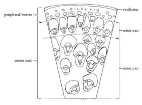
Figure 1. General structure and observation area (a), transversal section (b), lateral section (c)
Source: (Astuti, 1990; Krisdianto & Jasni, 2005; Mandang & Rulliaty, 2004)
c
Figure 1. General structure and observation area (a), transversal section (b), lateral section (c)
Source: (Astuti, 1990; Krisdianto & Jasni, 2005; Mandang & Rulliaty, 2004)
 a b
a b
 c d
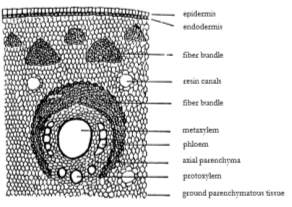
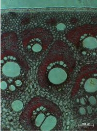
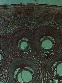
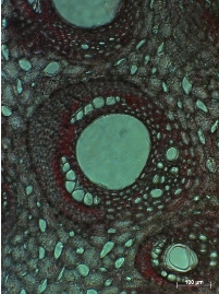
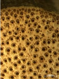
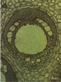
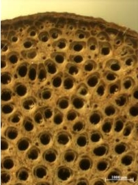
Figure 2. Anatomical structure of batu rattan stem (Calamus zonatus Beccari)
Remarks: a. Stem cross section, scale 1000 µm
b. Stem cross section, scale 100 µm
c. Single vascular bundle, scale 100 µm
d. Lateral section, scale 100 µm
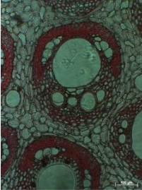
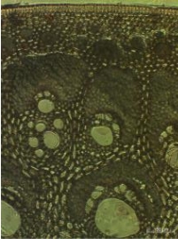
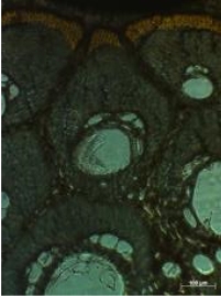
B. Buruk ati (Calamus insignis Griff. var. longispinosus Dransfield)
Epidermis is a single layer of unlignified parenchyma cells covered by siliceous interspersed with stomata cells. The epidermis layer is about 32.9 ± 3.78 µm and the endodermis layer is about 10.23 ± 2.23 µm. In the peripheral area, fiber bundles are arranged in one layer with no specific pattern (Figure 3b). The peripheral area is about 352.48 ± 61.66 µm from the outer bark. Vascular bundle in the outer part is about 7 – 8 per mm2, with the width and length (W/L) ratio is 0.9 ± 0.2, which means vascular bundle’s width is approximately similar with vascular bundle’s length. Yellow cap is absent, and metaxyem vessel diameter is 107.42 ± 31.83 µm.
Similar with those of monocotyledonous stem structure, vascular bundle in the inner part is greater in diameter than those of the outer part. The round to oblique vascular bundle length and width is 338.35 ± 68.49 µm and 296.59 ± 52.32 µm, respectively. The vascular bundle in the inner part is only 2 – 4 per mm2, with the width and length (W/L) ratio is 1.05 ± 0.14. Metaxylem vessels are single, with diameter of 295.1 ± 8.47 µm. Protoxylem consists of a cluster 2 – 4 cells, with single cell diameter of 37.9 ± 8.04 µm. Phloem double stranded fields lying laterally to the metaxylem vessels and every field contains 4 – 6 cells (Figure 3c). Individual phloem cell diameter is about 46.83 ± 8.73 µm. Resin canals are not found, intercellular spaces found in ground parenchyma tissue.
Fiber sheath is extensively longer in vascular bundle of peripheral area and normal ‘horse-shoe shaped’ in vascular bundle of inner part. Fiber length is 2,381.07 ± 246.98 µm, width 22 ± 2.52 µm, lumen 12.09 ± 2.73 µm and wall thickness 4.95 ± 0.66 µm. Ground parenchyma round to oval with varying sizes, type A.
c d
Figure 2. Anatomical structure of batu rattan stem (Calamus zonatus Beccari)
Remarks: a. Stem cross section, scale 1000 µm
b. Stem cross section, scale 100 µm
c. Single vascular bundle, scale 100 µm
d. Lateral section, scale 100 µm
B. Buruk ati (Calamus insignis Griff. var. longispinosus Dransfield)
Epidermis is a single layer of unlignified parenchyma cells covered by siliceous interspersed with stomata cells. The epidermis layer is about 32.9 ± 3.78 µm and the endodermis layer is about 10.23 ± 2.23 µm. In the peripheral area, fiber bundles are arranged in one layer with no specific pattern (Figure 3b). The peripheral area is about 352.48 ± 61.66 µm from the outer bark. Vascular bundle in the outer part is about 7 – 8 per mm2, with the width and length (W/L) ratio is 0.9 ± 0.2, which means vascular bundle’s width is approximately similar with vascular bundle’s length. Yellow cap is absent, and metaxyem vessel diameter is 107.42 ± 31.83 µm.
Similar with those of monocotyledonous stem structure, vascular bundle in the inner part is greater in diameter than those of the outer part. The round to oblique vascular bundle length and width is 338.35 ± 68.49 µm and 296.59 ± 52.32 µm, respectively. The vascular bundle in the inner part is only 2 – 4 per mm2, with the width and length (W/L) ratio is 1.05 ± 0.14. Metaxylem vessels are single, with diameter of 295.1 ± 8.47 µm. Protoxylem consists of a cluster 2 – 4 cells, with single cell diameter of 37.9 ± 8.04 µm. Phloem double stranded fields lying laterally to the metaxylem vessels and every field contains 4 – 6 cells (Figure 3c). Individual phloem cell diameter is about 46.83 ± 8.73 µm. Resin canals are not found, intercellular spaces found in ground parenchyma tissue.
Fiber sheath is extensively longer in vascular bundle of peripheral area and normal ‘horse-shoe shaped’ in vascular bundle of inner part. Fiber length is 2,381.07 ± 246.98 µm, width 22 ± 2.52 µm, lumen 12.09 ± 2.73 µm and wall thickness 4.95 ± 0.66 µm. Ground parenchyma round to oval with varying sizes, type A.
 a b
a b
 c d
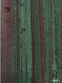
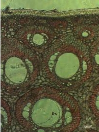
Figure 3. Anatomical structure of buruk ati rattan stem (Calamus insignis Griff. var. longispinosus Dransfield)
Remarks: a. Stem cross section, scale 1000 µm
b. Stem cross section, scale 100 µm
c. Single vascular bundle, scale 100 µm
d. Lateral section, scale 100 µm
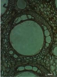
C. Duduk (Daemonorops didymophylla Beccari)
A single layer of unlignified parenchyma cells of epidermis layer covered by siliceous interspersed with few stomata cells. The epidermis layer is about 30.67 ± 2.77 µm and the endodermis layer is about 6.83 ± 1.59 µm. In the peripheral area, fiber bundles are arranged in one layer in alternate pattern, smaller and greater fiber bundles one after another (Figure 4b). The peripheral area is about 237.93 ± 18.62 µm from the outer bark. Vascular bundle in the outer part is about 9 – 11 per mm2, with the width and length (W/L) ratio is 0.72 ± 0.09, which means vascular bundle is approximately oval. Yellow cap is absent, and metaxyem vessel diameter is 104.76 ± 28.5 µm.
Vascular bundle in the inner part is greater in diameter than those of the outer part as commonly found in monocotyledonous stem. The oblique vascular bundle length is 451.18 ± 24.15 µm and width is 339.09 ± 57.24 µm. The vascular bundle in the inner part is 4 – 6 per mm2, with the width and length (W/L) ratio is 0.75 ± 0.11. Metaxylem vessels are single, with diameter of 176.16 ± 25.62 µm. Protoxylem consists of a cluster 2 – 5 cells, with single cell diameter of 26.51 ± 3.66 µm. Phloem double stranded fields lying laterally to the metaxylem vessels and every field contains 4 – 6 cells (Figure 4c). Individual phloem cell diameter is about 26.61 ± 3.5 µm. Resin canal diameter is 36.19 ± 10.01 µm.
Fiber sheath is mostly normal ‘horse-shoe shaped’ in inner part’s vascular bundle and some tapering in the outer part. Fiber length is 1,790.55 ± 228.42 µm, width 20.8 ± 1.17 µm, lumen 13.15 ± 0.99 µm and wall thickness 3.82 ± 0.37 µm. Ground parenchyma round to oval with varying sizes, type B.
.
c d
Figure 3. Anatomical structure of buruk ati rattan stem (Calamus insignis Griff. var. longispinosus Dransfield)
Remarks: a. Stem cross section, scale 1000 µm
b. Stem cross section, scale 100 µm
c. Single vascular bundle, scale 100 µm
d. Lateral section, scale 100 µm
C. Duduk (Daemonorops didymophylla Beccari)
A single layer of unlignified parenchyma cells of epidermis layer covered by siliceous interspersed with few stomata cells. The epidermis layer is about 30.67 ± 2.77 µm and the endodermis layer is about 6.83 ± 1.59 µm. In the peripheral area, fiber bundles are arranged in one layer in alternate pattern, smaller and greater fiber bundles one after another (Figure 4b). The peripheral area is about 237.93 ± 18.62 µm from the outer bark. Vascular bundle in the outer part is about 9 – 11 per mm2, with the width and length (W/L) ratio is 0.72 ± 0.09, which means vascular bundle is approximately oval. Yellow cap is absent, and metaxyem vessel diameter is 104.76 ± 28.5 µm.
Vascular bundle in the inner part is greater in diameter than those of the outer part as commonly found in monocotyledonous stem. The oblique vascular bundle length is 451.18 ± 24.15 µm and width is 339.09 ± 57.24 µm. The vascular bundle in the inner part is 4 – 6 per mm2, with the width and length (W/L) ratio is 0.75 ± 0.11. Metaxylem vessels are single, with diameter of 176.16 ± 25.62 µm. Protoxylem consists of a cluster 2 – 5 cells, with single cell diameter of 26.51 ± 3.66 µm. Phloem double stranded fields lying laterally to the metaxylem vessels and every field contains 4 – 6 cells (Figure 4c). Individual phloem cell diameter is about 26.61 ± 3.5 µm. Resin canal diameter is 36.19 ± 10.01 µm.
Fiber sheath is mostly normal ‘horse-shoe shaped’ in inner part’s vascular bundle and some tapering in the outer part. Fiber length is 1,790.55 ± 228.42 µm, width 20.8 ± 1.17 µm, lumen 13.15 ± 0.99 µm and wall thickness 3.82 ± 0.37 µm. Ground parenchyma round to oval with varying sizes, type B.
.
 a b
a b
 c d
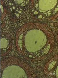
Figure 4. Anatomical structure of duduk rattan stem (Daemonorops didymophylla Beccari)
Remarks: a. Stem cross section, scale 1000 µm
b. Stem cross section, scale 100 µm
c. Single vascular bundle, scale 100 µm
d. Lateral section, scale 100 µm
D. Getah (Daemonorops micracantha (Griff.) Beccari)
Single layer epidermis consisting of unlignified parenchyma cells and covered by siliceous layer. The epidermis layer is about 29.93 ± 3.11 µm and the endodermis layer is about 12.84 ± 3.38 µm. In the peripheral area, fiber bundles are arranged in one layer with no specific pattern (Figure 5b). The peripheral area is about 208.74 ± 24.27 µm from the outer bark. Vascular bundle in the outer part is about 8 – 11 per mm2, with the width and length (W/L) ratio is 0.73 ± 0.11, which means vascular bundle is approximately oval. Yellow cap is absent, and metaxyem vessel diameter is 66.32 ± 12.26 µm.
As mostly found in monocotyledonous stem, vascular bundle in the inner part is greater in diameter than those of the outer part. The oval vascular bundle length and width is 533.25 ± 31.18 and 438.46 ± 74.25 µm, respectively. The vascular bundle in the inner part is 4 – 6 per mm2, with the width and length (W/L) ratio is 0.83 ± 0.18. Metaxylem vessels are single, with diameter of 250.88 ± 13.05 µm. Protoxylem consists of a cluster 4 – 6 cells, with single cell diameter of 57.78 ± 9.42 µm. Phloem double stranded fields lying laterally to the metaxylem vessels and every field contains of 4 – 6 cells (Figure 5c). Individual phloem cell diameter is about 50.31 ± 9.07 µm. Resin canal diameter is 100.23 ± 22.71 µm.
Fiber sheath is mostly normal ‘horse-shoe shaped’ in inner part’s vascular bundle and tapering in the outer part. Fiber length is 2,254.43 ± 292.55 µm, width 20.3 ± 2.54 µm, lumen 13.15 ± 2.74 µm and wall thickness 3.41 ± 0.47 µm. Ground parenchyma round to oval with varying sizes, type B.
c d
Figure 4. Anatomical structure of duduk rattan stem (Daemonorops didymophylla Beccari)
Remarks: a. Stem cross section, scale 1000 µm
b. Stem cross section, scale 100 µm
c. Single vascular bundle, scale 100 µm
d. Lateral section, scale 100 µm
D. Getah (Daemonorops micracantha (Griff.) Beccari)
Single layer epidermis consisting of unlignified parenchyma cells and covered by siliceous layer. The epidermis layer is about 29.93 ± 3.11 µm and the endodermis layer is about 12.84 ± 3.38 µm. In the peripheral area, fiber bundles are arranged in one layer with no specific pattern (Figure 5b). The peripheral area is about 208.74 ± 24.27 µm from the outer bark. Vascular bundle in the outer part is about 8 – 11 per mm2, with the width and length (W/L) ratio is 0.73 ± 0.11, which means vascular bundle is approximately oval. Yellow cap is absent, and metaxyem vessel diameter is 66.32 ± 12.26 µm.
As mostly found in monocotyledonous stem, vascular bundle in the inner part is greater in diameter than those of the outer part. The oval vascular bundle length and width is 533.25 ± 31.18 and 438.46 ± 74.25 µm, respectively. The vascular bundle in the inner part is 4 – 6 per mm2, with the width and length (W/L) ratio is 0.83 ± 0.18. Metaxylem vessels are single, with diameter of 250.88 ± 13.05 µm. Protoxylem consists of a cluster 4 – 6 cells, with single cell diameter of 57.78 ± 9.42 µm. Phloem double stranded fields lying laterally to the metaxylem vessels and every field contains of 4 – 6 cells (Figure 5c). Individual phloem cell diameter is about 50.31 ± 9.07 µm. Resin canal diameter is 100.23 ± 22.71 µm.
Fiber sheath is mostly normal ‘horse-shoe shaped’ in inner part’s vascular bundle and tapering in the outer part. Fiber length is 2,254.43 ± 292.55 µm, width 20.3 ± 2.54 µm, lumen 13.15 ± 2.74 µm and wall thickness 3.41 ± 0.47 µm. Ground parenchyma round to oval with varying sizes, type B.
 a b
a b
 c d
Figure 5. Anatomical structure of getah rattan stem (Daemonorops micracantha (Griff.) Beccari)
Remarks: a. Stem cross section, scale 1000 µm
b. Stem cross section, scale 100 µm
c. Single vascular bundle, scale 100 µm
d. Lateral section, scale 100 µm
E. Opon (Plectocomiopsis geminiflora (Griff.) Beccari)
Epidermis is a single layer of unlignified parenchyma cells covered by siliceous interspersed with few stomata cells. The epidermis layer is about 30.93 ± 6.24 µm and the endodermis layer is about 4.81 ± 0.96 µm. In the peripheral area, fiber bundles are arranged in one layer with no specific pattern (Figure 6b). The peripheral area is about 145.67 ± 18.4 µm from the outer bark. Vascular bundle in the outer part is about 7 – 9 per mm2, with the width and length (W/L) ratio is 0.98 ± 0.39, which means vascular bundle is approximately round. Yellow cap is found on the top of outer fiber bundle. Metaxyem vessel diameter is 192.59 ± 77.84 µm.
Vascular bundle in the outer part is extensively longer and narrower in the outer part than those in the inner part. In the inner part, vascular bundle is 479.49 ± 87.28 µm length and 438.46 ± 74.25 µm width. The vascular bundle in the inner part is 6 – 8 per mm2, with the width and length (W/L) ratio is 0.88 ± 0.16. Metaxylem vessels are mostly single in the outer part, while pair metaxylem vessels are found in the inner part with diameter of 313.2 ± 18.13 µm. Protoxylem consists of a cluster 2 – 4 cells, with single cell diameter of 35.81 ± 11.97 µm. Phloem double stranded fields lying laterally to the metaxylem vessels and every field contains of 3 – 6 cells (Figure 6c). Individual phloem cell diameter is about 43.43 ± 6.9 µm. Resin canal diameter is 86.8 ± 20.4 µm.
Fiber sheath is mostly normal ‘horse-shoe shaped’ in inner part’s vascular bundle and tapering in the outer part. Fiber length is 3,518.51 ± 474.51 µm, width 18.1 ± 2.9 µm, lumen 12 ± 2.19 µm and wall thickness 3.05 ± 0.69 µm. Ground parenchyma round to oval with varying sizes, type B.
c d
Figure 5. Anatomical structure of getah rattan stem (Daemonorops micracantha (Griff.) Beccari)
Remarks: a. Stem cross section, scale 1000 µm
b. Stem cross section, scale 100 µm
c. Single vascular bundle, scale 100 µm
d. Lateral section, scale 100 µm
E. Opon (Plectocomiopsis geminiflora (Griff.) Beccari)
Epidermis is a single layer of unlignified parenchyma cells covered by siliceous interspersed with few stomata cells. The epidermis layer is about 30.93 ± 6.24 µm and the endodermis layer is about 4.81 ± 0.96 µm. In the peripheral area, fiber bundles are arranged in one layer with no specific pattern (Figure 6b). The peripheral area is about 145.67 ± 18.4 µm from the outer bark. Vascular bundle in the outer part is about 7 – 9 per mm2, with the width and length (W/L) ratio is 0.98 ± 0.39, which means vascular bundle is approximately round. Yellow cap is found on the top of outer fiber bundle. Metaxyem vessel diameter is 192.59 ± 77.84 µm.
Vascular bundle in the outer part is extensively longer and narrower in the outer part than those in the inner part. In the inner part, vascular bundle is 479.49 ± 87.28 µm length and 438.46 ± 74.25 µm width. The vascular bundle in the inner part is 6 – 8 per mm2, with the width and length (W/L) ratio is 0.88 ± 0.16. Metaxylem vessels are mostly single in the outer part, while pair metaxylem vessels are found in the inner part with diameter of 313.2 ± 18.13 µm. Protoxylem consists of a cluster 2 – 4 cells, with single cell diameter of 35.81 ± 11.97 µm. Phloem double stranded fields lying laterally to the metaxylem vessels and every field contains of 3 – 6 cells (Figure 6c). Individual phloem cell diameter is about 43.43 ± 6.9 µm. Resin canal diameter is 86.8 ± 20.4 µm.
Fiber sheath is mostly normal ‘horse-shoe shaped’ in inner part’s vascular bundle and tapering in the outer part. Fiber length is 3,518.51 ± 474.51 µm, width 18.1 ± 2.9 µm, lumen 12 ± 2.19 µm and wall thickness 3.05 ± 0.69 µm. Ground parenchyma round to oval with varying sizes, type B.
 a b
a b
 c d
Figure 6. Anatomical structure of opon rattan stem (Plectocomiopsis geminiflora (Griff.) Beccari)
Remarks: a. Stem cross section, scale 1000 µm
b. Stem cross section, scale 100 µm
c. Single vascular bundle, scale 100 µm
d. Lateral section, scale 100 µm
F. Paku (Calamus exillis Griff.)
Single layer epidermis consisting of unlignified parenchyma cells and covered by siliceous layer interspersed with few stomata cells. The epidermis layer is about 33.84 ± 2.27 µm and the endodermis layer is about 9.82 ± 0.97 µm. In the peripheral area, fiber bundles are arranged in one to two layers with no specific pattern (Figure 7b). The peripheral area is about 173.07 ± 15.46 µm from the outer bark. Vascular bundle in the outer part is about 14 – 15 per mm2, with the width and length (W/L) ratio is 1.05 ± 0.24, which means vascular bundle is approximately round with length and width ratio is about 1. Yellow cap is absent. Metaxyem vessel diameter is 48.69 ± 13.2 µm.
Vascular bundle’s shape in the outer part is approximately similar with those in the inner part. In the inner part, vascular bundle is 393.36 ± 35.71 µm length and 336.01 ± 49.33 µm width. The vascular bundle in the inner part is 7 – 11 per mm2, with the width and length (W/L) ratio is 0.86 ± 0.12. Metaxylem vessels are mostly single in the inner part, while pair metaxylem vessels sometime are found in the inner part. Vessel’s diameter is 167.31 ± 14.44 µm. Protoxylem consists of a cluster 2 – 4 cells, with single cell diameter of 33.54 ± 5.94 µm. Phloem double stranded fields lying laterally to the metaxylem vessels and every field contains of 4 – 6 cells (Figure 7c). Individual phloem cell diameter is about 26.22 ± 4.7 µm. Resin canal diameter is 34.51 ± 8.14 µm.
Fiber sheath is in ‘horse-shoe shaped’ and elongated both in inner and outer parts. Fiber length is 1,367.21 ± 199.23 µm, width 19.8 ± 2.48 µm, lumen 13.03 ± 2.08 µm and wall thickness 3.39 ± 0.64 µm. Ground parenchyma round to oval with varying sizes, type B.
c d
Figure 6. Anatomical structure of opon rattan stem (Plectocomiopsis geminiflora (Griff.) Beccari)
Remarks: a. Stem cross section, scale 1000 µm
b. Stem cross section, scale 100 µm
c. Single vascular bundle, scale 100 µm
d. Lateral section, scale 100 µm
F. Paku (Calamus exillis Griff.)
Single layer epidermis consisting of unlignified parenchyma cells and covered by siliceous layer interspersed with few stomata cells. The epidermis layer is about 33.84 ± 2.27 µm and the endodermis layer is about 9.82 ± 0.97 µm. In the peripheral area, fiber bundles are arranged in one to two layers with no specific pattern (Figure 7b). The peripheral area is about 173.07 ± 15.46 µm from the outer bark. Vascular bundle in the outer part is about 14 – 15 per mm2, with the width and length (W/L) ratio is 1.05 ± 0.24, which means vascular bundle is approximately round with length and width ratio is about 1. Yellow cap is absent. Metaxyem vessel diameter is 48.69 ± 13.2 µm.
Vascular bundle’s shape in the outer part is approximately similar with those in the inner part. In the inner part, vascular bundle is 393.36 ± 35.71 µm length and 336.01 ± 49.33 µm width. The vascular bundle in the inner part is 7 – 11 per mm2, with the width and length (W/L) ratio is 0.86 ± 0.12. Metaxylem vessels are mostly single in the inner part, while pair metaxylem vessels sometime are found in the inner part. Vessel’s diameter is 167.31 ± 14.44 µm. Protoxylem consists of a cluster 2 – 4 cells, with single cell diameter of 33.54 ± 5.94 µm. Phloem double stranded fields lying laterally to the metaxylem vessels and every field contains of 4 – 6 cells (Figure 7c). Individual phloem cell diameter is about 26.22 ± 4.7 µm. Resin canal diameter is 34.51 ± 8.14 µm.
Fiber sheath is in ‘horse-shoe shaped’ and elongated both in inner and outer parts. Fiber length is 1,367.21 ± 199.23 µm, width 19.8 ± 2.48 µm, lumen 13.03 ± 2.08 µm and wall thickness 3.39 ± 0.64 µm. Ground parenchyma round to oval with varying sizes, type B.
 a b
a b
 c d
Figure 7. Anatomical structure of paku rattan stem (Calamus exillis Griff.).
Remarks: a. Stem cross section, scale 1000 µm
b. Stem cross section, scale 100 µm
c. Single vascular bundle, scale 100 µm
d. Lateral section, scale 100 µm
G. Sijau (Calamus exillis Griff.)
A single layer of unlignified parenchyma cells of epidermis layer covered by siliceous interspersed with few stomata cells. The epidermis layer is about 55.35 ± 1.33 µm and the endodermis layer is about 10.67 ± 0.8 µm. In the peripheral area, fiber bundles are arranged in one layer pattern (Figure 8b). The peripheral area is about 259.48 ± 51.42 µm from the outer bark. Vascular bundle in the outer part is about 4 – 5 per mm2, with the width and length (W/L) ratio is 1.2 ± 0.38, which means vascular bundle is oblique widely. Yellow cap is absent, and metaxyem vessel diameter is 138.9 ± 38.13 µm.
Vascular bundle in the inner part is greater in diameter than those of the outer part as commonly found in monocotyledonous stem. In the inner part, the vascular bundle length is 590.82 ± 66.57 µm and width is 518.52 ± 56.19 µm. The vascular bundle in the inner part is 3 – 4 per mm2, with the width and length (W/L) ratio is 0.88 ± 0.11. Metaxylem vessels are single, with diameter of 255.54 ± 49.19 µm. Protoxylem consists of a cluster 2 – 4 cells, with single cell diameter of 40.88 ± 14.5 µm. Phloem double stranded fields lying laterally to the metaxylem vessels and every field contains 4 – 6 cells (Figure 8c). Individual phloem cell diameter is about 41.88 ± 6.82 µm. Resin canal is distributed evenly in the ground parenchymatous tissue togteher with intercellular spaces.
Fiber sheath is mostly elongated in outer part and normal ‘horse-shoe shaped’ in inner part’s vascular bundle. Fiber length is 1,958.94 ± 177.12 µm, width 21.9 ± 3.39 µm, lumen 12.92 ± 2.75 µm and wall thickness 4.49 ± 0.74 µm. Ground parenchyma round to oval with varying sizes, type B.
c d
Figure 7. Anatomical structure of paku rattan stem (Calamus exillis Griff.).
Remarks: a. Stem cross section, scale 1000 µm
b. Stem cross section, scale 100 µm
c. Single vascular bundle, scale 100 µm
d. Lateral section, scale 100 µm
G. Sijau (Calamus exillis Griff.)
A single layer of unlignified parenchyma cells of epidermis layer covered by siliceous interspersed with few stomata cells. The epidermis layer is about 55.35 ± 1.33 µm and the endodermis layer is about 10.67 ± 0.8 µm. In the peripheral area, fiber bundles are arranged in one layer pattern (Figure 8b). The peripheral area is about 259.48 ± 51.42 µm from the outer bark. Vascular bundle in the outer part is about 4 – 5 per mm2, with the width and length (W/L) ratio is 1.2 ± 0.38, which means vascular bundle is oblique widely. Yellow cap is absent, and metaxyem vessel diameter is 138.9 ± 38.13 µm.
Vascular bundle in the inner part is greater in diameter than those of the outer part as commonly found in monocotyledonous stem. In the inner part, the vascular bundle length is 590.82 ± 66.57 µm and width is 518.52 ± 56.19 µm. The vascular bundle in the inner part is 3 – 4 per mm2, with the width and length (W/L) ratio is 0.88 ± 0.11. Metaxylem vessels are single, with diameter of 255.54 ± 49.19 µm. Protoxylem consists of a cluster 2 – 4 cells, with single cell diameter of 40.88 ± 14.5 µm. Phloem double stranded fields lying laterally to the metaxylem vessels and every field contains 4 – 6 cells (Figure 8c). Individual phloem cell diameter is about 41.88 ± 6.82 µm. Resin canal is distributed evenly in the ground parenchymatous tissue togteher with intercellular spaces.
Fiber sheath is mostly elongated in outer part and normal ‘horse-shoe shaped’ in inner part’s vascular bundle. Fiber length is 1,958.94 ± 177.12 µm, width 21.9 ± 3.39 µm, lumen 12.92 ± 2.75 µm and wall thickness 4.49 ± 0.74 µm. Ground parenchyma round to oval with varying sizes, type B.
 a b
a b
 c d
Figure 8. Anatomical structure of sijau rattan stem (Calamus laevigatus Martius)
Remarks: a. Stem cross section, scale 1000 µm
b. Stem cross section, scale 100 µm
c. Single vascular bundle, scale 100 µm
d. Lateral section, scale 100 µm
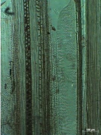
H. Tunggal (Calamus laevigatus Martius)
Single layer epidermis consisting of unlignified parenchyma cells and covered by siliceous layer. The epidermis layer is about 36.89 ± 4.06 µm and the endodermis layer is about 6.42 ± 1.1 µm. In the peripheral area, fiber bundles are arranged in one layer with no specific pattern (Figure 9b). The peripheral area is about 117.94 ± 13.3 µm from the outer bark. Vascular bundle in the outer part is about 4 – 5 per mm2, with the width and length (W/L) ratio is 0.95 ± 0.17, which means vascular bundle is approximately round. Yellow cap is absent, and metaxyem vessel diameter is 114.49 ± 30.84 µm.
As mostly found in monocotyledonous stem, vascular bundle in the inner part is greater in diameter than those of the outer part. The round vascular bundle length and width is 443.7 ± 27.06 and 436.3 ± 29.32 µm, respectively. The vascular bundle in the inner part is 4 – 5 per mm2, with the width and length (W/L) ratio is 0.99 ± 0.09. Metaxylem vessels are single, with diameter of 226.76 ± 28.46 µm. Protoxylem consists of a cluster 3 – 5 cells, with single cell diameter of 37.96 ± 5.17 µm. Phloem double stranded fields lying laterally to the metaxylem vessels and every field contains of 4 – 6 cells (Figure 9c). Individual phloem cell diameter is about 49.62 ± 7.21 µm. Resin canal is distributed evenly in the ground parenchymatous tissue togteher with intercellular spaces.
Fiber sheath is mostly normal ‘horse-shoe shaped’ in inner and outer parts. Fiber length is 2,126.57 ± 252.84 µm, width 19 ± 2.89 µm, lumen 10.59 ± 2.69 µm and wall thickness 4.2 ± 0.71 µm. Ground parenchyma round to oval with varying sizes, type A.
c d
Figure 8. Anatomical structure of sijau rattan stem (Calamus laevigatus Martius)
Remarks: a. Stem cross section, scale 1000 µm
b. Stem cross section, scale 100 µm
c. Single vascular bundle, scale 100 µm
d. Lateral section, scale 100 µm
H. Tunggal (Calamus laevigatus Martius)
Single layer epidermis consisting of unlignified parenchyma cells and covered by siliceous layer. The epidermis layer is about 36.89 ± 4.06 µm and the endodermis layer is about 6.42 ± 1.1 µm. In the peripheral area, fiber bundles are arranged in one layer with no specific pattern (Figure 9b). The peripheral area is about 117.94 ± 13.3 µm from the outer bark. Vascular bundle in the outer part is about 4 – 5 per mm2, with the width and length (W/L) ratio is 0.95 ± 0.17, which means vascular bundle is approximately round. Yellow cap is absent, and metaxyem vessel diameter is 114.49 ± 30.84 µm.
As mostly found in monocotyledonous stem, vascular bundle in the inner part is greater in diameter than those of the outer part. The round vascular bundle length and width is 443.7 ± 27.06 and 436.3 ± 29.32 µm, respectively. The vascular bundle in the inner part is 4 – 5 per mm2, with the width and length (W/L) ratio is 0.99 ± 0.09. Metaxylem vessels are single, with diameter of 226.76 ± 28.46 µm. Protoxylem consists of a cluster 3 – 5 cells, with single cell diameter of 37.96 ± 5.17 µm. Phloem double stranded fields lying laterally to the metaxylem vessels and every field contains of 4 – 6 cells (Figure 9c). Individual phloem cell diameter is about 49.62 ± 7.21 µm. Resin canal is distributed evenly in the ground parenchymatous tissue togteher with intercellular spaces.
Fiber sheath is mostly normal ‘horse-shoe shaped’ in inner and outer parts. Fiber length is 2,126.57 ± 252.84 µm, width 19 ± 2.89 µm, lumen 10.59 ± 2.69 µm and wall thickness 4.2 ± 0.71 µm. Ground parenchyma round to oval with varying sizes, type A.
 a b
a b
 c d
Figure 9. Anatomical structure of tunggal rattan stem (Calamus laevigatus Martius)
Remarks: a. Stem cross section, scale 1000 µm
b. Stem cross section, scale 100 µm
c. Single vascular bundle, scale 100 µm
d. Lateral section, scale 100 µm
I. Udang (Korthalsia flagelaris Miquel)
Single layer epidermis consisting of unlignified parenchyma cells and covered by siliceous layer. The epidermis layer is about 63.39 ± 6.25 µm and the endodermis layer is about 10.34 ± 4.43 µm. In the peripheral area, fiber bundles are arranged in one layer with no specific pattern. The peripheral area is about 208.67 ± 12.77 µm from the outer bark. Vascular bundle in the outer part is about 7 – 8 per mm2, with the width and length (W/L) ratio is 0.97 ± 0.41, which means vascular bundle is approximately oval. Yellow cap is found in the top of outer vascular bundle, metaxyem vessel diameter is 142.81 ± 35.79 µm.
As mostly found in monocotyledonous stem, vascular bundle in the inner part is greater in diameter than those of the outer part. The oval vascular bundle length and width is 673.72 ± 76.96 and 480.26 ± 68.17 µm, respectively. The vascular bundle in the inner part is 6 – 7 per mm2, with the width and length (W/L) ratio is 0.73 ± 0.18. Metaxylem vessels are single, with diameter of 319.02 ± 33.74 µm. Protoxylem consists of a cluster 4 – 6 cells, with single cell diameter of 57.05 ± 5.81 µm. Phloem double stranded fields lying laterally to the metaxylem vessels and every field contains of 4 – 6 cells (Figure 10c). Individual phloem cell diameter is about 51.52 ± 8.87 µm. Resin canal diameter is 100.43 ± 15.84 µm.
Fiber sheath is mostly normal ‘horse-shoe shaped’ in inner part’s vascular bundle and elongated in the outer part. Fiber length is 2,476.86 ± 332.08 µm, width 23.3 ± 2.8 µm, lumen 13.57 ± 1.94 µm and wall thickness 4.86 ± 1.05 µm. Ground parenchyma round to oval with varying sizes, type B.
c d
Figure 9. Anatomical structure of tunggal rattan stem (Calamus laevigatus Martius)
Remarks: a. Stem cross section, scale 1000 µm
b. Stem cross section, scale 100 µm
c. Single vascular bundle, scale 100 µm
d. Lateral section, scale 100 µm
I. Udang (Korthalsia flagelaris Miquel)
Single layer epidermis consisting of unlignified parenchyma cells and covered by siliceous layer. The epidermis layer is about 63.39 ± 6.25 µm and the endodermis layer is about 10.34 ± 4.43 µm. In the peripheral area, fiber bundles are arranged in one layer with no specific pattern. The peripheral area is about 208.67 ± 12.77 µm from the outer bark. Vascular bundle in the outer part is about 7 – 8 per mm2, with the width and length (W/L) ratio is 0.97 ± 0.41, which means vascular bundle is approximately oval. Yellow cap is found in the top of outer vascular bundle, metaxyem vessel diameter is 142.81 ± 35.79 µm.
As mostly found in monocotyledonous stem, vascular bundle in the inner part is greater in diameter than those of the outer part. The oval vascular bundle length and width is 673.72 ± 76.96 and 480.26 ± 68.17 µm, respectively. The vascular bundle in the inner part is 6 – 7 per mm2, with the width and length (W/L) ratio is 0.73 ± 0.18. Metaxylem vessels are single, with diameter of 319.02 ± 33.74 µm. Protoxylem consists of a cluster 4 – 6 cells, with single cell diameter of 57.05 ± 5.81 µm. Phloem double stranded fields lying laterally to the metaxylem vessels and every field contains of 4 – 6 cells (Figure 10c). Individual phloem cell diameter is about 51.52 ± 8.87 µm. Resin canal diameter is 100.43 ± 15.84 µm.
Fiber sheath is mostly normal ‘horse-shoe shaped’ in inner part’s vascular bundle and elongated in the outer part. Fiber length is 2,476.86 ± 332.08 µm, width 23.3 ± 2.8 µm, lumen 13.57 ± 1.94 µm and wall thickness 4.86 ± 1.05 µm. Ground parenchyma round to oval with varying sizes, type B.
 a b
a b
 c d
Figure 10. Anatomical structure of udang rattan stem (Korthalsia flagelaris Miquel)
Remarks: a. Stem cross section, scale 1000 µm
b. Stem cross section, scale 100 µm
c. Single vascular bundle, scale 100 µm
d. Lateral section, scale 100 µm
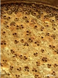
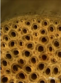
As generally found in monocotyledonous, stem comprise a large central core of primary vascular bundles embedded in parenchymatous ground tissue, surrounded by peripheral area/cortex (Butterfield & Meylan, 1980). The general stem anatomy of species investigated is similar with some Indonesian rattan species described by Jasni, Damayanti, and Kalima, (2012); Jasni, Damayanti, Kalima, Malik, and Abdurachman, (2010); Jasni, Krisdianto, Kalima, and Abdurachman (2012). All species investigated were distinctly made up of epidermis, cortex and central cylinder. All species exhibited a single layer epidermis consisting of unlignified parenchyma cells. The epidermis was covered with siliceous layer interspersed with few stomata cells. The cortex or known as peripheral area consisted of fiber bands, undeveloped vascular bundles embedded in parenchyma cells of varying shapes and sizes structured ring-like in the outer part. Yellow cap which covers the top part of outer vascular bundle is found in opon (Plectocomiopsis geminiflora (Griff.) Beccari) (Figure 6b) and udang (Korthalsia flagelaris Miquel) (Figure 10b).
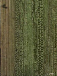
The central cylinder of all species was generally composed of vascular bundles of varying sizes and structure embedded in ground parenchyma. The vascular bundle consisted of conducting tissue (xylem and phloem), which was surrounded by fiber sheath and parenchyma. The xylem was mostly composed of one large metaxylem-vessel and a cluster of protoxylem-vessels. In some species investigated, tyloses-like was found in few metaxylem-vessels. The phloem consisted of different number of sieve tubes with companion cells. Ground parenchyma round to oval with varying sizes, type A and B, as shown in longitudinal section appear like ‘stacks of coins’. Tentative identification key to rattan species originated from Jambi is shown below.
c d
Figure 10. Anatomical structure of udang rattan stem (Korthalsia flagelaris Miquel)
Remarks: a. Stem cross section, scale 1000 µm
b. Stem cross section, scale 100 µm
c. Single vascular bundle, scale 100 µm
d. Lateral section, scale 100 µm
As generally found in monocotyledonous, stem comprise a large central core of primary vascular bundles embedded in parenchymatous ground tissue, surrounded by peripheral area/cortex (Butterfield & Meylan, 1980). The general stem anatomy of species investigated is similar with some Indonesian rattan species described by Jasni, Damayanti, and Kalima, (2012); Jasni, Damayanti, Kalima, Malik, and Abdurachman, (2010); Jasni, Krisdianto, Kalima, and Abdurachman (2012). All species investigated were distinctly made up of epidermis, cortex and central cylinder. All species exhibited a single layer epidermis consisting of unlignified parenchyma cells. The epidermis was covered with siliceous layer interspersed with few stomata cells. The cortex or known as peripheral area consisted of fiber bands, undeveloped vascular bundles embedded in parenchyma cells of varying shapes and sizes structured ring-like in the outer part. Yellow cap which covers the top part of outer vascular bundle is found in opon (Plectocomiopsis geminiflora (Griff.) Beccari) (Figure 6b) and udang (Korthalsia flagelaris Miquel) (Figure 10b).
The central cylinder of all species was generally composed of vascular bundles of varying sizes and structure embedded in ground parenchyma. The vascular bundle consisted of conducting tissue (xylem and phloem), which was surrounded by fiber sheath and parenchyma. The xylem was mostly composed of one large metaxylem-vessel and a cluster of protoxylem-vessels. In some species investigated, tyloses-like was found in few metaxylem-vessels. The phloem consisted of different number of sieve tubes with companion cells. Ground parenchyma round to oval with varying sizes, type A and B, as shown in longitudinal section appear like ‘stacks of coins’. Tentative identification key to rattan species originated from Jambi is shown below.
II. MATERIAL AND METHOD
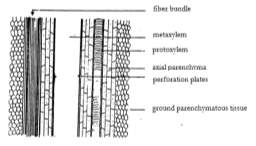
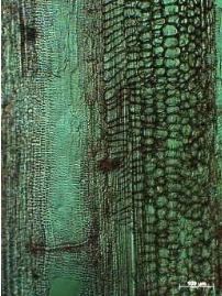
Nine rattan species are collected from Marangin-Bangko forest, Jambi Province, where nine indigenous rattan species grows naturally in the area. Nine matured stems were collected randomly from different growing clumps for each species. Herbarium samples were collected for species identification purposes in the Botany Research Group, Forest Research and Development Center (P3H), while rattan stem were collected for macroscopic, microscopic, and fiber dimension assessments. Macroscopically, solid rattan stem was cut using sharp knife and was observed under stereo-microscope. Microscopically, cross-sectional rattan stems were sliced into 35 – 45 µm using a microtome. These sectioned were then red-stained using safranin, dehydrated in ethanol sequence (35 – 100%) prior to mount it in object glass with entellan. (Sass, 1961 as cited by Rulliaty, 2014). Two milimetre by two milimetre transversal stem in two centimetres length stem was macerated according to modified Forest Products Laboratory method as mentioned by Rulliaty (2014). Rattan stem observation area is modified based on Astuti (1990), Krisdianto and Jasni (2005), and Jasni, Krisdianto, Kalima, and Abdurachman, (2012). Rattan stem anatomical properties is observed based on Mandang and Rulliaty (2004), Weiner and Liese (1990) and Krisdianto and Jasni (2005). The schematic observation is shown in Figure 1a, and anatomical rattan stem properties is shown in Figure 1b and 1c.
a b
c
Figure 1. General structure and observation area (a), transversal section (b), lateral section (c)
Source: (Astuti, 1990; Krisdianto & Jasni, 2005; Mandang & Rulliaty, 2004)
III. RESULT AND DISCUSSION
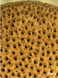
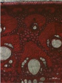
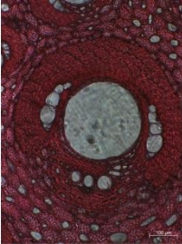
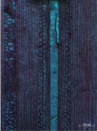
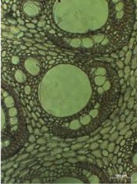
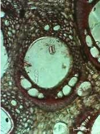
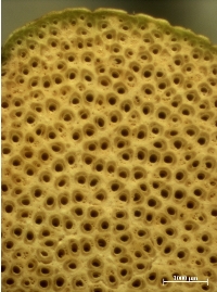
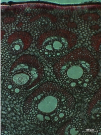
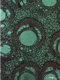
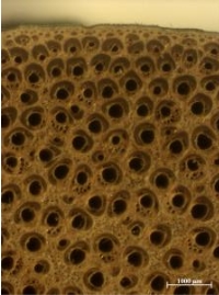
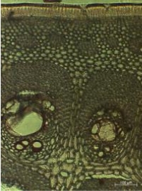
Anatomical properties of nine rattan species are described as follows: A. Batu (Calamus zonatus Beccari) Single layer epidermis consisting of unlignified parenchyma cells and covered by siliceous layer interspersed with few stomata cells. The epidermis layer is about 19.01 ± 1.92 µm and the endodermis layer is about 4.6 ± 0.75 µm. In the peripheral area, fiber bundles are arranged in one to two layers with resin canals found in the peripheral area (Figure 2b). The peripheral area is about 136.81 ± 9.26 µm from the outer bark. Vascular bundle in the outer part is about 12 – 13 per mm2, with the width and length (W/L) ratio is 0.9 ± 0.3, which means vascular bundle’s width is approximately similar with vascular bundle’s length. Yellow cap is absent, and metaxyem vessel diameter is 99.27 ± 27.22 µm. As commonly found in monocotyledonous stem structure, vascular bundle in the inner part is greater in diameter than those of the outer part. The elipse to oblique vascular bundle length and width is 448.4 ± 53.6 µm and 359.75 ± 43.04 µm, respectively. The vascular bundle in the inner part is 6 – 8 per mm2, with the width and length (W/L) ratio is 0.81 ± 0.08. Metaxylem vessels are mostly single, sometime found in pair, with diameter of 211.35 ± 26.65 µm. Protoxylem consists of a cluster 2 – 6 cells, with single cell diameter of 40.61 ± 10.65 µm. Phloem double stranded fields lying laterally to the metaxylem vessels and every field contains 4 – 6 cells (Figure 2c). Individual phloem cell diameter is about 27.89 ± 6.17 µm. Resin canals are diffusely distributed in the inner, outer and peripheral areas. Resin canal diameter is 68.81 ± 10.23 µm. Fiber sheath is extensively broader in vascular bundle of peripheral area and normal ‘horse-shoe shaped’ in vascular bundle of inner part. Fiber length is 1,821.16 ± 169 µm, width 20.4 ± 3.21 µm, lumen 11.76 ± 2.57 µm and wall thickness 4.31 ± 0.71 µm. Ground parenchyma round to oval with varying sizes, type A.
a b
c d
Figure 2. Anatomical structure of batu rattan stem (Calamus zonatus Beccari)
Remarks: a. Stem cross section, scale 1000 µm
b. Stem cross section, scale 100 µm
c. Single vascular bundle, scale 100 µm
d. Lateral section, scale 100 µm
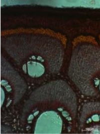
B. Buruk ati (Calamus insignis Griff. var. longispinosus Dransfield)
Epidermis is a single layer of unlignified parenchyma cells covered by siliceous interspersed with stomata cells. The epidermis layer is about 32.9 ± 3.78 µm and the endodermis layer is about 10.23 ± 2.23 µm. In the peripheral area, fiber bundles are arranged in one layer with no specific pattern (Figure 3b). The peripheral area is about 352.48 ± 61.66 µm from the outer bark. Vascular bundle in the outer part is about 7 – 8 per mm2, with the width and length (W/L) ratio is 0.9 ± 0.2, which means vascular bundle’s width is approximately similar with vascular bundle’s length. Yellow cap is absent, and metaxyem vessel diameter is 107.42 ± 31.83 µm.
Similar with those of monocotyledonous stem structure, vascular bundle in the inner part is greater in diameter than those of the outer part. The round to oblique vascular bundle length and width is 338.35 ± 68.49 µm and 296.59 ± 52.32 µm, respectively. The vascular bundle in the inner part is only 2 – 4 per mm2, with the width and length (W/L) ratio is 1.05 ± 0.14. Metaxylem vessels are single, with diameter of 295.1 ± 8.47 µm. Protoxylem consists of a cluster 2 – 4 cells, with single cell diameter of 37.9 ± 8.04 µm. Phloem double stranded fields lying laterally to the metaxylem vessels and every field contains 4 – 6 cells (Figure 3c). Individual phloem cell diameter is about 46.83 ± 8.73 µm. Resin canals are not found, intercellular spaces found in ground parenchyma tissue.
Fiber sheath is extensively longer in vascular bundle of peripheral area and normal ‘horse-shoe shaped’ in vascular bundle of inner part. Fiber length is 2,381.07 ± 246.98 µm, width 22 ± 2.52 µm, lumen 12.09 ± 2.73 µm and wall thickness 4.95 ± 0.66 µm. Ground parenchyma round to oval with varying sizes, type A.
a b
c d
Figure 3. Anatomical structure of buruk ati rattan stem (Calamus insignis Griff. var. longispinosus Dransfield)
Remarks: a. Stem cross section, scale 1000 µm
b. Stem cross section, scale 100 µm
c. Single vascular bundle, scale 100 µm
d. Lateral section, scale 100 µm
C. Duduk (Daemonorops didymophylla Beccari)
A single layer of unlignified parenchyma cells of epidermis layer covered by siliceous interspersed with few stomata cells. The epidermis layer is about 30.67 ± 2.77 µm and the endodermis layer is about 6.83 ± 1.59 µm. In the peripheral area, fiber bundles are arranged in one layer in alternate pattern, smaller and greater fiber bundles one after another (Figure 4b). The peripheral area is about 237.93 ± 18.62 µm from the outer bark. Vascular bundle in the outer part is about 9 – 11 per mm2, with the width and length (W/L) ratio is 0.72 ± 0.09, which means vascular bundle is approximately oval. Yellow cap is absent, and metaxyem vessel diameter is 104.76 ± 28.5 µm.
Vascular bundle in the inner part is greater in diameter than those of the outer part as commonly found in monocotyledonous stem. The oblique vascular bundle length is 451.18 ± 24.15 µm and width is 339.09 ± 57.24 µm. The vascular bundle in the inner part is 4 – 6 per mm2, with the width and length (W/L) ratio is 0.75 ± 0.11. Metaxylem vessels are single, with diameter of 176.16 ± 25.62 µm. Protoxylem consists of a cluster 2 – 5 cells, with single cell diameter of 26.51 ± 3.66 µm. Phloem double stranded fields lying laterally to the metaxylem vessels and every field contains 4 – 6 cells (Figure 4c). Individual phloem cell diameter is about 26.61 ± 3.5 µm. Resin canal diameter is 36.19 ± 10.01 µm.
Fiber sheath is mostly normal ‘horse-shoe shaped’ in inner part’s vascular bundle and some tapering in the outer part. Fiber length is 1,790.55 ± 228.42 µm, width 20.8 ± 1.17 µm, lumen 13.15 ± 0.99 µm and wall thickness 3.82 ± 0.37 µm. Ground parenchyma round to oval with varying sizes, type B.
.
a b
c d
Figure 4. Anatomical structure of duduk rattan stem (Daemonorops didymophylla Beccari)
Remarks: a. Stem cross section, scale 1000 µm
b. Stem cross section, scale 100 µm
c. Single vascular bundle, scale 100 µm
d. Lateral section, scale 100 µm
D. Getah (Daemonorops micracantha (Griff.) Beccari)
Single layer epidermis consisting of unlignified parenchyma cells and covered by siliceous layer. The epidermis layer is about 29.93 ± 3.11 µm and the endodermis layer is about 12.84 ± 3.38 µm. In the peripheral area, fiber bundles are arranged in one layer with no specific pattern (Figure 5b). The peripheral area is about 208.74 ± 24.27 µm from the outer bark. Vascular bundle in the outer part is about 8 – 11 per mm2, with the width and length (W/L) ratio is 0.73 ± 0.11, which means vascular bundle is approximately oval. Yellow cap is absent, and metaxyem vessel diameter is 66.32 ± 12.26 µm.
As mostly found in monocotyledonous stem, vascular bundle in the inner part is greater in diameter than those of the outer part. The oval vascular bundle length and width is 533.25 ± 31.18 and 438.46 ± 74.25 µm, respectively. The vascular bundle in the inner part is 4 – 6 per mm2, with the width and length (W/L) ratio is 0.83 ± 0.18. Metaxylem vessels are single, with diameter of 250.88 ± 13.05 µm. Protoxylem consists of a cluster 4 – 6 cells, with single cell diameter of 57.78 ± 9.42 µm. Phloem double stranded fields lying laterally to the metaxylem vessels and every field contains of 4 – 6 cells (Figure 5c). Individual phloem cell diameter is about 50.31 ± 9.07 µm. Resin canal diameter is 100.23 ± 22.71 µm.
Fiber sheath is mostly normal ‘horse-shoe shaped’ in inner part’s vascular bundle and tapering in the outer part. Fiber length is 2,254.43 ± 292.55 µm, width 20.3 ± 2.54 µm, lumen 13.15 ± 2.74 µm and wall thickness 3.41 ± 0.47 µm. Ground parenchyma round to oval with varying sizes, type B.
a b
c d
Figure 5. Anatomical structure of getah rattan stem (Daemonorops micracantha (Griff.) Beccari)
Remarks: a. Stem cross section, scale 1000 µm
b. Stem cross section, scale 100 µm
c. Single vascular bundle, scale 100 µm
d. Lateral section, scale 100 µm
E. Opon (Plectocomiopsis geminiflora (Griff.) Beccari)
Epidermis is a single layer of unlignified parenchyma cells covered by siliceous interspersed with few stomata cells. The epidermis layer is about 30.93 ± 6.24 µm and the endodermis layer is about 4.81 ± 0.96 µm. In the peripheral area, fiber bundles are arranged in one layer with no specific pattern (Figure 6b). The peripheral area is about 145.67 ± 18.4 µm from the outer bark. Vascular bundle in the outer part is about 7 – 9 per mm2, with the width and length (W/L) ratio is 0.98 ± 0.39, which means vascular bundle is approximately round. Yellow cap is found on the top of outer fiber bundle. Metaxyem vessel diameter is 192.59 ± 77.84 µm.
Vascular bundle in the outer part is extensively longer and narrower in the outer part than those in the inner part. In the inner part, vascular bundle is 479.49 ± 87.28 µm length and 438.46 ± 74.25 µm width. The vascular bundle in the inner part is 6 – 8 per mm2, with the width and length (W/L) ratio is 0.88 ± 0.16. Metaxylem vessels are mostly single in the outer part, while pair metaxylem vessels are found in the inner part with diameter of 313.2 ± 18.13 µm. Protoxylem consists of a cluster 2 – 4 cells, with single cell diameter of 35.81 ± 11.97 µm. Phloem double stranded fields lying laterally to the metaxylem vessels and every field contains of 3 – 6 cells (Figure 6c). Individual phloem cell diameter is about 43.43 ± 6.9 µm. Resin canal diameter is 86.8 ± 20.4 µm.
Fiber sheath is mostly normal ‘horse-shoe shaped’ in inner part’s vascular bundle and tapering in the outer part. Fiber length is 3,518.51 ± 474.51 µm, width 18.1 ± 2.9 µm, lumen 12 ± 2.19 µm and wall thickness 3.05 ± 0.69 µm. Ground parenchyma round to oval with varying sizes, type B.
a b
c d
Figure 6. Anatomical structure of opon rattan stem (Plectocomiopsis geminiflora (Griff.) Beccari)
Remarks: a. Stem cross section, scale 1000 µm
b. Stem cross section, scale 100 µm
c. Single vascular bundle, scale 100 µm
d. Lateral section, scale 100 µm
F. Paku (Calamus exillis Griff.)
Single layer epidermis consisting of unlignified parenchyma cells and covered by siliceous layer interspersed with few stomata cells. The epidermis layer is about 33.84 ± 2.27 µm and the endodermis layer is about 9.82 ± 0.97 µm. In the peripheral area, fiber bundles are arranged in one to two layers with no specific pattern (Figure 7b). The peripheral area is about 173.07 ± 15.46 µm from the outer bark. Vascular bundle in the outer part is about 14 – 15 per mm2, with the width and length (W/L) ratio is 1.05 ± 0.24, which means vascular bundle is approximately round with length and width ratio is about 1. Yellow cap is absent. Metaxyem vessel diameter is 48.69 ± 13.2 µm.
Vascular bundle’s shape in the outer part is approximately similar with those in the inner part. In the inner part, vascular bundle is 393.36 ± 35.71 µm length and 336.01 ± 49.33 µm width. The vascular bundle in the inner part is 7 – 11 per mm2, with the width and length (W/L) ratio is 0.86 ± 0.12. Metaxylem vessels are mostly single in the inner part, while pair metaxylem vessels sometime are found in the inner part. Vessel’s diameter is 167.31 ± 14.44 µm. Protoxylem consists of a cluster 2 – 4 cells, with single cell diameter of 33.54 ± 5.94 µm. Phloem double stranded fields lying laterally to the metaxylem vessels and every field contains of 4 – 6 cells (Figure 7c). Individual phloem cell diameter is about 26.22 ± 4.7 µm. Resin canal diameter is 34.51 ± 8.14 µm.
Fiber sheath is in ‘horse-shoe shaped’ and elongated both in inner and outer parts. Fiber length is 1,367.21 ± 199.23 µm, width 19.8 ± 2.48 µm, lumen 13.03 ± 2.08 µm and wall thickness 3.39 ± 0.64 µm. Ground parenchyma round to oval with varying sizes, type B.
a b
c d
Figure 7. Anatomical structure of paku rattan stem (Calamus exillis Griff.).
Remarks: a. Stem cross section, scale 1000 µm
b. Stem cross section, scale 100 µm
c. Single vascular bundle, scale 100 µm
d. Lateral section, scale 100 µm
G. Sijau (Calamus exillis Griff.)
A single layer of unlignified parenchyma cells of epidermis layer covered by siliceous interspersed with few stomata cells. The epidermis layer is about 55.35 ± 1.33 µm and the endodermis layer is about 10.67 ± 0.8 µm. In the peripheral area, fiber bundles are arranged in one layer pattern (Figure 8b). The peripheral area is about 259.48 ± 51.42 µm from the outer bark. Vascular bundle in the outer part is about 4 – 5 per mm2, with the width and length (W/L) ratio is 1.2 ± 0.38, which means vascular bundle is oblique widely. Yellow cap is absent, and metaxyem vessel diameter is 138.9 ± 38.13 µm.
Vascular bundle in the inner part is greater in diameter than those of the outer part as commonly found in monocotyledonous stem. In the inner part, the vascular bundle length is 590.82 ± 66.57 µm and width is 518.52 ± 56.19 µm. The vascular bundle in the inner part is 3 – 4 per mm2, with the width and length (W/L) ratio is 0.88 ± 0.11. Metaxylem vessels are single, with diameter of 255.54 ± 49.19 µm. Protoxylem consists of a cluster 2 – 4 cells, with single cell diameter of 40.88 ± 14.5 µm. Phloem double stranded fields lying laterally to the metaxylem vessels and every field contains 4 – 6 cells (Figure 8c). Individual phloem cell diameter is about 41.88 ± 6.82 µm. Resin canal is distributed evenly in the ground parenchymatous tissue togteher with intercellular spaces.
Fiber sheath is mostly elongated in outer part and normal ‘horse-shoe shaped’ in inner part’s vascular bundle. Fiber length is 1,958.94 ± 177.12 µm, width 21.9 ± 3.39 µm, lumen 12.92 ± 2.75 µm and wall thickness 4.49 ± 0.74 µm. Ground parenchyma round to oval with varying sizes, type B.
a b
c d
Figure 8. Anatomical structure of sijau rattan stem (Calamus laevigatus Martius)
Remarks: a. Stem cross section, scale 1000 µm
b. Stem cross section, scale 100 µm
c. Single vascular bundle, scale 100 µm
d. Lateral section, scale 100 µm
H. Tunggal (Calamus laevigatus Martius)
Single layer epidermis consisting of unlignified parenchyma cells and covered by siliceous layer. The epidermis layer is about 36.89 ± 4.06 µm and the endodermis layer is about 6.42 ± 1.1 µm. In the peripheral area, fiber bundles are arranged in one layer with no specific pattern (Figure 9b). The peripheral area is about 117.94 ± 13.3 µm from the outer bark. Vascular bundle in the outer part is about 4 – 5 per mm2, with the width and length (W/L) ratio is 0.95 ± 0.17, which means vascular bundle is approximately round. Yellow cap is absent, and metaxyem vessel diameter is 114.49 ± 30.84 µm.
As mostly found in monocotyledonous stem, vascular bundle in the inner part is greater in diameter than those of the outer part. The round vascular bundle length and width is 443.7 ± 27.06 and 436.3 ± 29.32 µm, respectively. The vascular bundle in the inner part is 4 – 5 per mm2, with the width and length (W/L) ratio is 0.99 ± 0.09. Metaxylem vessels are single, with diameter of 226.76 ± 28.46 µm. Protoxylem consists of a cluster 3 – 5 cells, with single cell diameter of 37.96 ± 5.17 µm. Phloem double stranded fields lying laterally to the metaxylem vessels and every field contains of 4 – 6 cells (Figure 9c). Individual phloem cell diameter is about 49.62 ± 7.21 µm. Resin canal is distributed evenly in the ground parenchymatous tissue togteher with intercellular spaces.
Fiber sheath is mostly normal ‘horse-shoe shaped’ in inner and outer parts. Fiber length is 2,126.57 ± 252.84 µm, width 19 ± 2.89 µm, lumen 10.59 ± 2.69 µm and wall thickness 4.2 ± 0.71 µm. Ground parenchyma round to oval with varying sizes, type A.
a b
c d
Figure 9. Anatomical structure of tunggal rattan stem (Calamus laevigatus Martius)
Remarks: a. Stem cross section, scale 1000 µm
b. Stem cross section, scale 100 µm
c. Single vascular bundle, scale 100 µm
d. Lateral section, scale 100 µm
I. Udang (Korthalsia flagelaris Miquel)
Single layer epidermis consisting of unlignified parenchyma cells and covered by siliceous layer. The epidermis layer is about 63.39 ± 6.25 µm and the endodermis layer is about 10.34 ± 4.43 µm. In the peripheral area, fiber bundles are arranged in one layer with no specific pattern. The peripheral area is about 208.67 ± 12.77 µm from the outer bark. Vascular bundle in the outer part is about 7 – 8 per mm2, with the width and length (W/L) ratio is 0.97 ± 0.41, which means vascular bundle is approximately oval. Yellow cap is found in the top of outer vascular bundle, metaxyem vessel diameter is 142.81 ± 35.79 µm.
As mostly found in monocotyledonous stem, vascular bundle in the inner part is greater in diameter than those of the outer part. The oval vascular bundle length and width is 673.72 ± 76.96 and 480.26 ± 68.17 µm, respectively. The vascular bundle in the inner part is 6 – 7 per mm2, with the width and length (W/L) ratio is 0.73 ± 0.18. Metaxylem vessels are single, with diameter of 319.02 ± 33.74 µm. Protoxylem consists of a cluster 4 – 6 cells, with single cell diameter of 57.05 ± 5.81 µm. Phloem double stranded fields lying laterally to the metaxylem vessels and every field contains of 4 – 6 cells (Figure 10c). Individual phloem cell diameter is about 51.52 ± 8.87 µm. Resin canal diameter is 100.43 ± 15.84 µm.
Fiber sheath is mostly normal ‘horse-shoe shaped’ in inner part’s vascular bundle and elongated in the outer part. Fiber length is 2,476.86 ± 332.08 µm, width 23.3 ± 2.8 µm, lumen 13.57 ± 1.94 µm and wall thickness 4.86 ± 1.05 µm. Ground parenchyma round to oval with varying sizes, type B.
a b
c d
Figure 10. Anatomical structure of udang rattan stem (Korthalsia flagelaris Miquel)
Remarks: a. Stem cross section, scale 1000 µm
b. Stem cross section, scale 100 µm
c. Single vascular bundle, scale 100 µm
d. Lateral section, scale 100 µm
As generally found in monocotyledonous, stem comprise a large central core of primary vascular bundles embedded in parenchymatous ground tissue, surrounded by peripheral area/cortex (Butterfield & Meylan, 1980). The general stem anatomy of species investigated is similar with some Indonesian rattan species described by Jasni, Damayanti, and Kalima, (2012); Jasni, Damayanti, Kalima, Malik, and Abdurachman, (2010); Jasni, Krisdianto, Kalima, and Abdurachman (2012). All species investigated were distinctly made up of epidermis, cortex and central cylinder. All species exhibited a single layer epidermis consisting of unlignified parenchyma cells. The epidermis was covered with siliceous layer interspersed with few stomata cells. The cortex or known as peripheral area consisted of fiber bands, undeveloped vascular bundles embedded in parenchyma cells of varying shapes and sizes structured ring-like in the outer part. Yellow cap which covers the top part of outer vascular bundle is found in opon (Plectocomiopsis geminiflora (Griff.) Beccari) (Figure 6b) and udang (Korthalsia flagelaris Miquel) (Figure 10b).
The central cylinder of all species was generally composed of vascular bundles of varying sizes and structure embedded in ground parenchyma. The vascular bundle consisted of conducting tissue (xylem and phloem), which was surrounded by fiber sheath and parenchyma. The xylem was mostly composed of one large metaxylem-vessel and a cluster of protoxylem-vessels. In some species investigated, tyloses-like was found in few metaxylem-vessels. The phloem consisted of different number of sieve tubes with companion cells. Ground parenchyma round to oval with varying sizes, type A and B, as shown in longitudinal section appear like ‘stacks of coins’. Tentative identification key to rattan species originated from Jambi is shown below.
- Vascular bundles in the outer part of the stem
- yellow capped ................................................................................................................. 2
- yellow cap absent ............................................................................................................ 3
- Metaxylem vessels in the inner part
- mostly in pair .................................................opon (Plectocomiopsis geminiflora (Griff.) Beccari).
- mostly single .............................................udang (Korthalsia flagelaris Miquel).
- Circle resin canals
- found in ground parenchymatous tissue ........................................................................... 4
- not found ............................................................................................................................... 7
- Resin canals
- in some area are found in pair ................. getah (Daemonorops micracantha (Griff.) Beccari)
- exclusively single ............................................................................................................. 5
- Single resin canals
- are found in center part and in cortex/peripheral area ....... batu (Calamus zonatus Beccari)
- are found in center part ..................................................................................................... 6
- Fiber bundles in the peripheral area
- is structured in one to two lines with no specific pattern .......... paku (Calamus exillis Griff.)
- is structured in one line with alternate pattern .... duduk (Daemonorops didymophylla Beccari)
- Fiber bundles in the central part
- are embedded in the ground parenchymatous tissue in between vascular bundle ....................................................................................... tunggal (Calamus laevigatus Martius)
- are not found separately from vascular bundle ................................................................. 8
- Vascular bundles in the outer part
- are round to oval with W/L ratio is more than 1 .............. sijau (Calamus laevigatus Martius)
- are oval to elongated with W/L ratio is less than 1 ....................................... buruk ati (Calamus insignis Griff. var. longispinosus Dransfield)
IV. conclusion
Nine rattan species originated from Jambi were anatomically described for species identification purposes. The anatomical properties could become a diagnostic characteristic for rattan species identification and specific characteristic has been developed for key species determination. Tentative identification key to rattan species has been developed for nine species investigated, then the key should be developed for further genera identification among rattan species in Indonesia.REFERENCES
Abasolo, W. P. (2015). Properties of rattan cane as basis for determining optimum cutting cycle of cultivated Calamus merrillii. Journal of Tropical Forest Science, 27(2), 176–188. Astuti, S. (1990). Anatomi perbandingan batang beberapa jenis rotan dari Karangkamulyan dan Pananjung Pangandaran, Jawa Barat. Institut Teknologi Bandung, Bandung. Butterfield, B. G., & Meylan, B. A. (1980). Three-dimensional structure of wood. London: Chapman and Hall. Ebanyenle, E., & Oteng-Amoako, A. A. (2003). Anatomy and identification of five indigenous rattan species of Ghana. Ghana Journal Forestry, 11(2), 77–90. Jasni, -, Damayanti, R., & Kalima, T. (2012). Atlas rotan Indonesia (Jilid 1, C). Bogor: Pusat Penelitian dan Pengembangan Keteknikan Kehutanan dan Pengolahan Hasil Hutan. Jasni, -, Damayanti, R., Kalima, T., Malik, J., & Abdurachman, -. (2010). Atlas rotan Indonesia (Jilid 2). Bogor: Pusat Penelitian dan Pengembangan Keteknikan Kehutanan dan Pengolahan Hasil Hutan. Jasni, -, Krisdianto, -, Kalima, T., & Abdurachman, -. (2012). Atlas rotan Indonesia (Jilid 3). Bogor: Pusat Penelitian dan Pengembangan Keteknikan Kehutanan dan Pengolahan Hasil Hutan. Krisdianto, -, & Jasni, -. (2005). Struktur anatomi tiga jenis batang rotan. Jurnal Ilmu Dan Teknologi Kayu Tropis, 3(2), 45–52. Mandang, Y. I., & Rulliaty, S. (2004). Anatomi batang rotan (No. 02.A-PEN-II-6). Bogor. Otniel, T. (2013, June 19). Rattan export ban makes its farmers miserable. Antaranews.com. Jakarta. Retrieved from http://www.antaranews.com/en/news/89443/rattan-export-ban-makes-its-farmers-miserable Rachman, O., & Jasni, -. (2013). Rotan - Sumber daya, sifat, dan pengolahannya. Bogor: Pusat Penelitian dan Pengembangan Keteknikan Kehutanan dan Pengolahan Hasil Hutan. Rulliaty, S. (2014). Identifikasi dan kualitas serat lima jenis kayu andalan setempat asal Jawa Barat dan Banten. Jurnal Penelitian Hasil Hutan, 32(4), 297–312. Sosef, M. S. M., Hong, L. T., & Prawirohatmodjo, S. (1998). Plant Resources of South East Asia 5(3) Timber trees: Lesser-known timbers. Bogor: PROSEA. Weiner, G., & Liese, W. (1990). Rattans - Stem anatomy and taxonomic implications. IAWA Bulletin, 11(1), 61–70. Zehui, J. (2007). Bamboo and rattan in the world. Beijing: China Forestry Publishing House.Cite This Work
To export a reference to this article please select a referencing stye below:
Reference Copied to Clipboard.
Reference Copied to Clipboard.
Reference Copied to Clipboard.
Reference Copied to Clipboard.
Reference Copied to Clipboard.
Reference Copied to Clipboard.
Reference Copied to Clipboard.
Related Services
View all


Related Content
All TagsContent relating to: "International Studies"
International Studies relates to the studying of economics, politics, culture, and other aspects of life on an international scale. International Studies allows you to develop an understanding of international relations and gives you an insight into global issues.
Related Articles

DMCA / Removal Request
If you are the original writer of this dissertation and no longer wish to have your work published on the UKDiss.com website then please: